

Macrophage Migration Inhibitory Factor (MIF) and Its Homologue d-Dopachrome Tautomerase (DDT) Inversely Correlate with Inflammation in Discoid Lupus Erythematosus
- PMID: 33401503
- PMCID: PMC7795694
- DOI: 10.3390/molecules26010184
Macrophage Migration Inhibitory Factor (MIF) and Its Homologue d-Dopachrome Tautomerase (DDT) Inversely Correlate with Inflammation in Discoid Lupus Erythematosus
Abstract
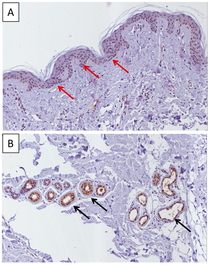
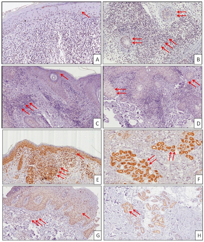
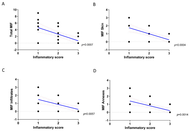
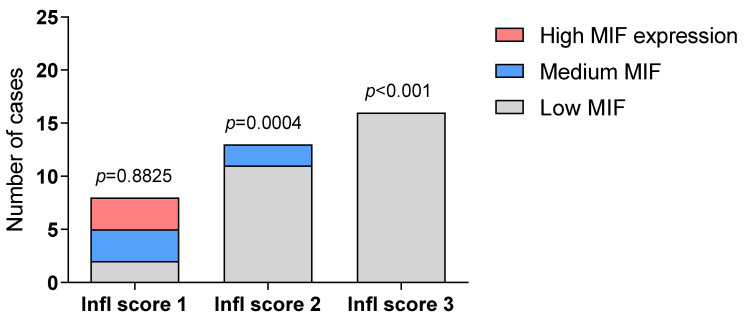
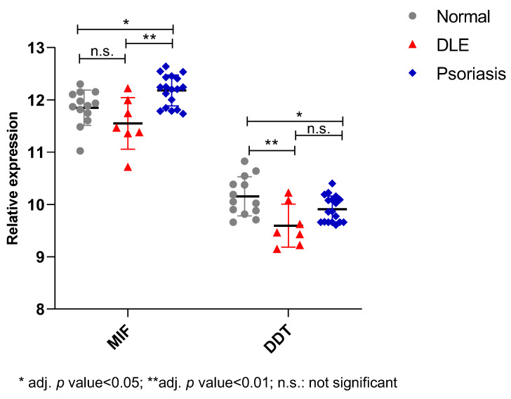
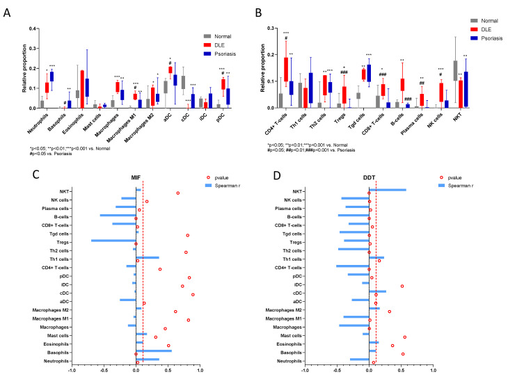
Discoid Lupus Erythematosus (DLE) is a chronic cutaneous disease of unknown etiology and of immunoinflammatory origin that is characterized by inflammatory plaques and may lead to disfiguring scarring and skin atrophy. Current treatments are limited, with a large proportion of patients either poorly or not responsive, which makes DLE an unmet medical need. Macrophage migration inhibitory factor (MIF) is the prototype of a pleiotropic family of cytokine that also includes the recently discovered homologue D-dopachrome tautomerase (DDT) or MIF2. MIF and DDT/MIF-2 exert several biological properties, primarily, but not exclusively of a proinflammatory nature. MIF and DDT have been suggested to play a key role in the pathogenesis of several autoimmune diseases, such as multiple sclerosis and type 1 diabetes, as well as in the development and progression of certain forms of cancers. In the present study, we have performed an immunohistochemistry analysis for the evaluation of MIF in DLE lesions and normal skin. We found high levels of MIF in the basal layer of the epidermis as well as in the cutaneous appendage (eccrine glands and sebocytes) of normal skin. In DLE lesions, we observed a significant negative correlation between the expression of MIF and the severity of inflammation. In addition, we performed an analysis of MIF and DDT expression levels in the skin of DLE patients in a publicly available microarray dataset. Interestingly, while these in silico data only evidenced a trend toward reduced levels of MIF, they demonstrated a significant pattern of expression and correlation of DDT with inflammatory infiltrates in DLE skins. Overall, our data support a protective role for endogenous MIF and possibly DDT in the regulation of homeostasis and inflammation in the skin and open up novel avenues for the treatment of DLE.
Keywords: d-dopachrome tautomerase; discoid lupus erythematosus; macrophage migration inhibitory factor.
Conflict of interest statement
The authors declare no conflict of interest.
Figures
Similar articles
-
Upregulated Expression of Macrophage Migration Inhibitory Factor, Its Analogue D-Dopachrome Tautomerase, and the CD44 Receptor in Peripheral CD4 T Cells from Clinically Isolated Syndrome Patients with Rapid Conversion to Clinical Defined Multiple Sclerosis.Medicina (Kaunas). 2019 Oct 1;55(10):667. doi: 10.3390/medicina55100667. Medicina (Kaunas). 2019. PMID: 31581595 Free PMC article.
-
UVB-induced inflammation gives increased d-dopachrome tautomerase activity in blister fluid which correlates with macrophage migration inhibitory factor.Exp Dermatol. 2003 Jun;12(3):278-82. doi: 10.1034/j.1600-0625.2003.120307.x. Exp Dermatol. 2003. PMID: 12823441
-
D-dopachrome tautomerase is over-expressed in pancreatic ductal adenocarcinoma and acts cooperatively with macrophage migration inhibitory factor to promote cancer growth.Int J Cancer. 2016 Nov 1;139(9):2056-67. doi: 10.1002/ijc.30278. Epub 2016 Jul 28. Int J Cancer. 2016. PMID: 27434219
-
The Role of Macrophage Migration Inhibitory Factor and Its Homolog D-Dopachrome Tautomerase in Ultraviolet Radiation-Induced Carcinogenesis: New Insights Into Skin Cancer Mechanisms.Photodermatol Photoimmunol Photomed. 2025 Sep;41(5):e70046. doi: 10.1111/phpp.70046. Photodermatol Photoimmunol Photomed. 2025. PMID: 40847634 Review.
-
Immune modulation by the macrophage migration inhibitory factor (MIF) family: D-dopachrome tautomerase (DDT) is not (always) a backup system.Cytokine. 2020 Sep;133:155121. doi: 10.1016/j.cyto.2020.155121. Epub 2020 May 11. Cytokine. 2020. PMID: 32417648 Review.
Cited by
-
Evaluation of the Involvement of Heme Oxygenase-1 Expression in Discoid Lupus Erythematosus Lesions.Antioxidants (Basel). 2023 Jun 27;12(7):1352. doi: 10.3390/antiox12071352. Antioxidants (Basel). 2023. PMID: 37507892 Free PMC article.
-
Serum and urinary levels of MIF, CD74, DDT and CXCR4 among patients with type 1 diabetes mellitus, type 2 diabetes and healthy individuals: Implications for further research.Metabol Open. 2024 Sep 15;24:100320. doi: 10.1016/j.metop.2024.100320. eCollection 2024 Dec. Metabol Open. 2024. PMID: 39323959 Free PMC article.
-
The Role of Macrophage Migration Inhibitory Factor (MIF) and D-Dopachrome Tautomerase (D-DT/MIF-2) in Infections: A Clinical Perspective.Biomedicines. 2023 Dec 19;12(1):2. doi: 10.3390/biomedicines12010002. Biomedicines. 2023. PMID: 38275363 Free PMC article. Review.
-
The mosaic of autoimmunity - Finally discussing in person. The 13th international congress on autoimmunity 2022 (AUTO13) Athens.Autoimmun Rev. 2022 Oct;21(10):103166. doi: 10.1016/j.autrev.2022.103166. Epub 2022 Aug 4. Autoimmun Rev. 2022. PMID: 35932955 Free PMC article.
-
Involvement of the Macrophage Migration Inhibitory Factor (MIF) in Lipedema.Metabolites. 2023 Oct 23;13(10):1105. doi: 10.3390/metabo13101105. Metabolites. 2023. PMID: 37887430 Free PMC article.
References
Publication types
MeSH terms
Substances
LinkOut - more resources
Full Text Sources
Other Literature Sources
Medical
Molecular Biology Databases
Miscellaneous
