. 2021 Jan 1;22(1):400.
doi: 10.3390/ijms22010400.

Crucial Role of FABP3 in αSyn-Induced Reduction of Septal GABAergic Neurons and Cognitive Decline in Mice

Affiliations

Crucial Role of FABP3 in αSyn-Induced Reduction of Septal GABAergic Neurons and Cognitive Decline in Mice

Kazuya Matsuo et al. Int J Mol Sci. .

Abstract

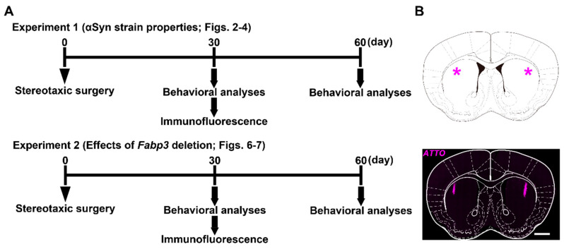
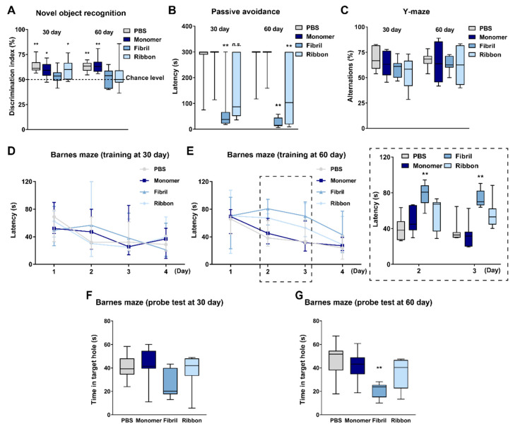
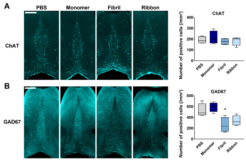
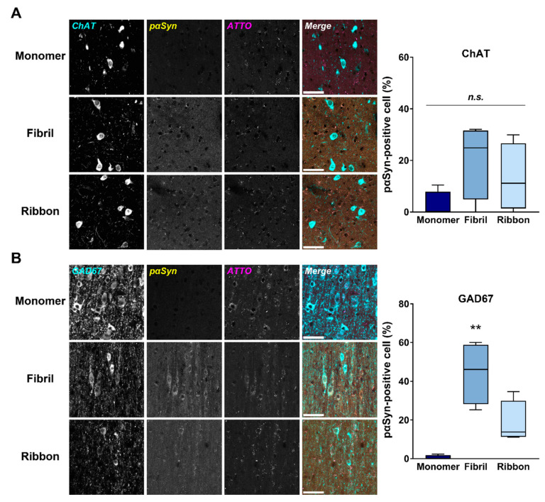
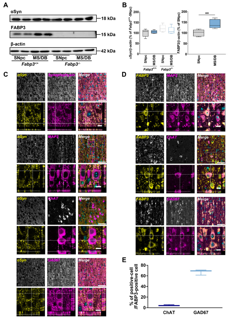
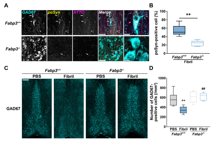
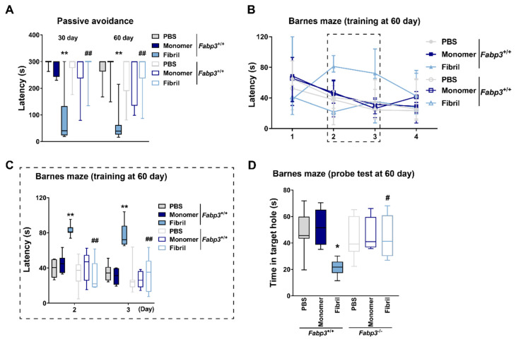
In synucleinopathies, while motor symptoms are thought to be attributed to the accumulation of misfolded α-synuclein (αSyn) in nigral dopaminergic neurons, it remains to be elucidated how cognitive decline arises. Here, we investigated the effects of distinct αSyn strains on cognition and the related neuropathology in the medial septum/diagonal band (MS/DB), a key region for cognitive processing. Bilateral injection of αSyn fibrils into the dorsal striatum potently impaired cognition in mice. The cognitive decline was accompanied by accumulation of phosphorylated αSyn at Ser129 and reduction of gamma-aminobutyric acid (GABA)-ergic but not cholinergic neurons in the MS/DB. Since we have demonstrated that fatty acid-binding protein 3 (FABP3) is critical for αSyn neurotoxicity in nigral dopaminergic neurons, we investigated whether FABP3 also participates in αSyn pathology in the MS/DB and cognitive decline. FABP3 was highly expressed in GABAergic but rarely in cholinergic neurons in the MS/DB. Notably, Fabp3 deletion antagonized the accumulation of phosphorylated αSyn, decrease in GABAergic neurons, and cognitive impairment caused by αSyn fibrils. Overall, the present study indicates that FABP3 mediates αSyn neurotoxicity in septal GABAergic neurons and the resultant cognitive impairment, and that FABP3 in this subpopulation could be a therapeutic target for dementia in synucleinopathies.

Keywords: cognition; fatty acid-binding protein; gamma-aminobutyric acid; medial septum; α-synuclein.


Conflict of interest statement

The authors declare no conflict of interest.

Figures

Figure 1
Figure 1
Experimental schedule and the injection site in mouse brain. (A) Experimental schedule in this study using intrastriatally α-synuclein (αSyn)-injected mice. Mice were divided into two groups, received the stereotaxic surgery, and were subjected to either Experiment 1 or 2. Experiment 1 was conducted to reveal αSyn strain-dependent properties regarding cognitive impairment and the related pathological alterations. Mice were injected with αSyn monomers, fibrils, or ribbons. Experiment 2 was conducted using fatty acid-binding protein 3 (Fabp3)−/− mice to address whether FABP3 participates in αSyn-induced cognitive impairment and the related pathology. Mice were injected with αSyn monomers or fibrils. (B) (Top) The injection site (*) is depicted. (Bottom) The actual site confirmed by direct visualization of immunoreactivity against ATTO550 (magenta). The image was obtained 30 days after the injection of αSyn fibrils and was overlaid on the diagram shown above. Scale bar: 1 mm.
Figure 2
Figure 2
αSyn strain-dependent cognitive impairments in intrastriatally injected mice. (A) Discrimination index in a novel object recognition task (n = 7 per group). * p < 0.05 and ** p < 0.01 between contacts with familiar and novel objects within groups. (B) Latency to enter the dark compartment in a passive avoidance task (n = 7 per group). ** p < 0.01 vs. PBS-injected group in the same day. n.s., not significant. (C) Spontaneous alternations in a Y-maze task (n = 7 per group). (D,E) Latency to enter the escape box in training sessions in a Barnes maze task (n = 7 per group). The sessions were conducted twice per day. The insertion depicts the days when the latency was significantly different between groups. ** p < 0.01 vs. PBS-injected group. (F,G) Latency to explore the target hole in probe test sessions in the Barnes maze task (n = 7 per group). The test was conducted 7 days after the first training sessions. ** p < 0.01 vs. PBS-injected group.
Figure 3
Figure 3
Selective decrease in septal GABAergic neurons after intrastriatal injection of αSyn. (Left) Representative images of choline acetyltransferase (ChAT)-positive (A) and glutamic acid decarboxylase 67 (GAD67)-positive (B) cells in the medial septum/diagonal band (MS/DB), 30 days after the injection. Scale bar: 500 μm. (Right) Quantitative analyses of each protein-positive cell number in the MS/DB. * p < 0.05 vs. PBS-injected group (n = 4 per group).
Figure 4
Figure 4
Accumulation of pαSyn in septal GABAergic neurons after intrastriatal injection of αSyn. (Left) Representative images of phosphorylated αSyn (pαSyn; yellow) accumulation and exogenously applied αSyn (ATTO; magenta) in ChAT-positive (A) and GAD67-positive (B) cells (cyan) in the MS/DB, 30 days after the injection. Scale bar: 50 μm. (Right) Quantitative analyses of the ratio of pαSyn-positive cells in ChAT-positive (A) and GAD67-positive cells. ** p < 0.01 vs. monomeric αSyn-injected group (n = 4 per group).
Figure 5
Figure 5
Properties of expression levels and the localization of endogenous αSyn and FABP3 in the MS/DB. (A) Representative images of blots probed with anti-αSyn and FABP3 antibodies detected in lysates of substantia nigra pars compacta (SNpc) and MS/DB from naïve Fabp3+/+ and Fabp3−/− mice. (B) Quantitative analyses of the blots. ** p < 0.01 between SNpc and MS/DB from Fabp3+/+ mice (n = 5 per group). (C,D) Representative images of the localization of endogenous αSyn (C) and FABP3 (D) in the MS/DB. Sections were probed with anti-αSyn (C) or anti-FABP3 (D) antibody (yellow) with anti-synaptophysin, microtubule-associated protein 2 (MAP2), ChAT, or GAD67 antibodies (magenta). The lower images of each staining represent the magnification and the orthogonal XZ and YZ planes. Scale bar: 50 (merge) and 10 (magnification) μm, respectively. Sections were counterstained with 4′,6-diamidino-2-phenylindole (cyan). (E) Quantitative analyses of the ratio of ChAT-positive or GAD67-positive cells in FABP3-positive cells in the MS/DB (n = 3 each).
Figure 6
Figure 6
Effects of Fabp3 deletion on pαSyn accumulation and reduction of septal GABAergic neurons after intrastriatal αSyn injection. (A) Representative images of pαSyn (yellow) accumulation in GAD67-positive cells (cyan) in the MS/DB, 30 days after the injection. Arrows and arrowheads depict pαSyn-accumulated GAD67-positive and -negative cells, respectively. Scale bar: 50 (merge) and 10 (magnification) μm, respectively. (B) Quantitative analyses of the ratio of pαSyn-positive cells in GAD67-positive cells (n = 6 per group). ** p < 0.01 between αSyn fibril-injected Fabp3+/+ and Fabp3−/− mice. (C) Representative images of GAD67-positive cells (cyan) in the MS/DB, 30 days after the injection. Scale bar: 250 μm. (D) Quantitative analyses of GAD67-positive cells (n = 6 per group). ** p < 0.01 vs. PBS-injected Fabp3+/+ mice; ## p < 0.01 vs. αSyn fibril-injected Fabp3+/+ mice.
Figure 7
Figure 7
Effects of Fabp3 deletion on cognitive impairments after intrastriatal αSyn injection. (A) Latency to enter the dark box in the passive avoidance task (n = 7 per group). ** p < 0.01 vs. PBS-injected Fabp3+/+ mice and ## p < 0.01 vs. αSyn fibril-injected Fabp3+/+ mice in the same day. (B,C) Latency to enter the escape box in training sessions in the Barnes maze task (n = 7 per group). The sessions were conducted twice per day. The insertion (C) depicts the days when the latency was significantly different between groups. ** p < 0.01 vs. PBS-injected Fabp3+/+ mice; ## p < 0.01 vs. αSyn fibril-injected Fabp3+/+ mice. (D) Latency to explore the target hole in the probe test sessions in the Barnes maze task (n = 7 per group). The test was conducted 7 days after the first training sessions. * p < 0.05 vs. PBS-injected Fabp3+/+ mice. # p < 0.05 vs. αSyn fibril-injected Fabp3+/+ mice.

Similar articles

Cited by

References

    1. Spillantini M.G., Schmidt M.L., Lee V.M.Y., Trojanowski J.Q., Jakes R., Goedert M. α-synuclein in Lewy bodies. Nature. 1997;388:839–840. doi: 10.1038/42166. - DOI - PubMed
    1. Hughes A.J., Daniel S.E., Kilford L., Lees A.J. Accuracy of clinical diagnosis of idiopathic Parkinson’s disease: A clinico-pathological study of 100 cases. J. Neurol. Neurosurg. Psychiatry. 1992;55:181–184. doi: 10.1136/jnnp.55.3.181. - DOI - PMC - PubMed
    1. Buter T.C., van den Hout A., Matthews F.E., Larsen J.P., Brayne C., Aarsland D. Dementia and survival in Parkinson disease: A 12-year population study. Neurology. 2008;70:1017–1022. doi: 10.1212/01.wnl.0000306632.43729.24. - DOI - PubMed
    1. Hely M.A., Reid W.G.J., Adena M.A., Halliday G.M., Morris J.G.L. The Sydney multicenter study of Parkinson’s disease: The inevitability of dementia at 20 years. Mov. Disord. 2008;23:837–844. doi: 10.1002/mds.21956. - DOI - PubMed
    1. Mattila P.M., Rinne J.O., Helenius H., Dickson D.W., Röyttä M. Alpha-synuclein-immunoreactive cortical Lewy bodies are associated with cognitive impairment in Parkinson’s disease. Acta Neuropathol. 2000;100:285–290. doi: 10.1007/s004019900168. - DOI - PubMed

MeSH terms