. 2021 Jan 3;22(1):417.
doi: 10.3390/ijms22010417.

Durable Oral Biofilm Resistance of 3D-Printed Dental Base Polymers Containing Zwitterionic Materials

Affiliations

Durable Oral Biofilm Resistance of 3D-Printed Dental Base Polymers Containing Zwitterionic Materials

Jae-Sung Kwon et al. Int J Mol Sci. .

Abstract

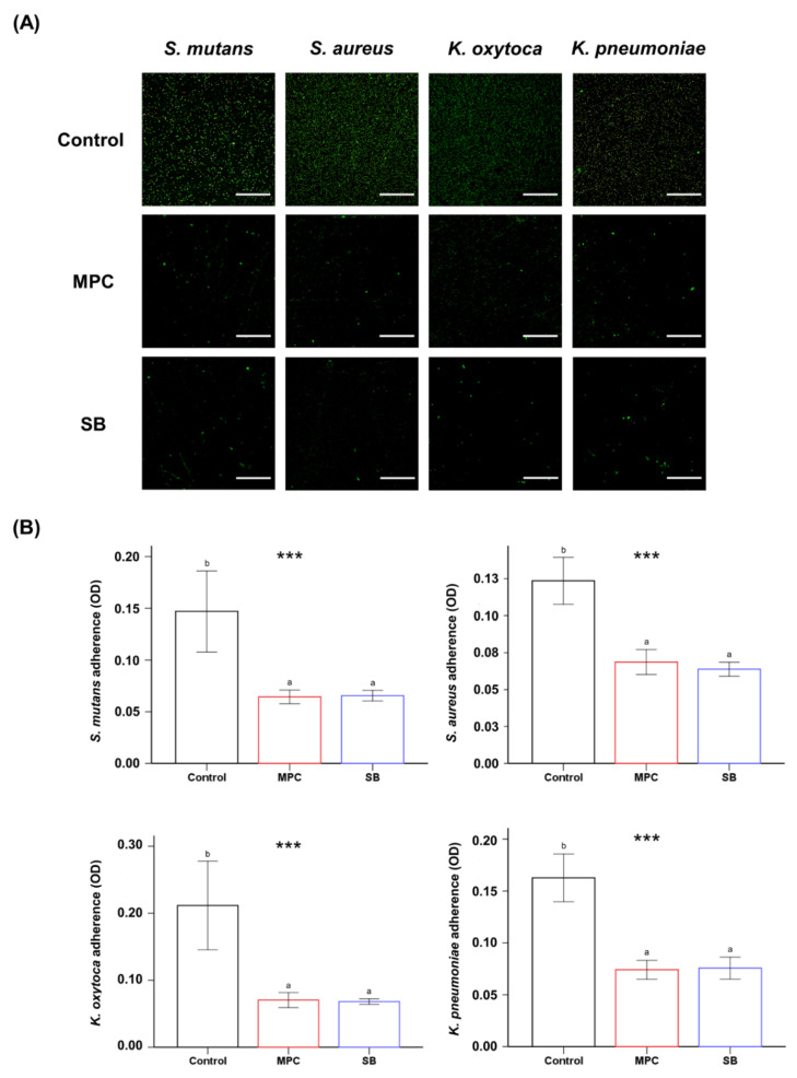
Poly(methyl methacralyate) (PMMA) has long been used in dentistry as a base polymer for dentures, and it is recently being used for the 3D printing of dental materials. Despite its many advantages, its susceptibility to microbial colonization remains to be overcome. In this study, the interface between 3D-printed PMMA specimens and oral salivary biofilm was studied following the addition of zwitterionic materials, 2-methacryloyloxyethyl phosphorylcholine (MPC) or sulfobetaine methacrylate (SB). A significant reduction in bacterial and biofilm adhesions was observed following the addition of MPC or SB, owing to their protein-repellent properties, and there were no significant differences between the two test materials. Although the mechanical properties of the tested materials were degraded, the statistical value of the reduction was minimal and all the properties fulfilled the requirements set by the International Standard, ISO 20795-2. Additionally, both the test materials maintained their resistance to biofilm when subjected to hydrothermal fatigue, with no further deterioration of the mechanical properties. Thus, novel 3D-printable PMMA incorporated with MPC or SB shows durable oral salivary biofilm resistance with maintenance of the physical and mechanical properties.

Keywords: 3D printing; dental base resin; dentistry; durability; oral salivary biofilm; poly(methyl methacralyate); zwitterion.


Conflict of interest statement

The authors declare no conflict of interest.

Figures

Figure 1
Figure 1
Physical and mechanical properties in terms of the contact angle (A,B), flexural strength (C,D), elastic modulus (E,F), and Vickers hardness (G,H) of poly(methyl methacralyate) (PMMA) before (Control) and after the addition of 3 wt% 2-methacryloyloxyethyl phosphorylcholine (MPC) or sulfobetaine methacrylate (SB). Results obtained before (left) and after thermocycling (right) are shown. Different lowercase letters above the bars indicate significant differences by post-hoc Tukey’s test. *** p < 0.001, ** p < 0.01, * p < 0.05 for comparisons between PMMA with and without the zwitterion analyzed by one-way analysis of variance (ANOVA).
Figure 2
Figure 2
Comparison of the optical density (OD) of the adsorbed bovine serum albumin (BSA) (A) and protein adsorbed from the brain heart infusion (BHI) medium (B) of poly(methyl methacralyate) PMMA samples before (Control) and after the addition of 2-methacryloyloxyethyl phosphorylcholine (MPC) or sulfobetaine methacrylate (SB). Different lowercase letters above bars indicate significant differences by post-hoc Tukey’s test. *** p < 0.001 for comparisons between PMMA with and without the zwitterion analyzed by one-way analysis of variance (ANOVA).
Figure 3
Figure 3
(A) Live and dead images of four different bacteria (S. mutans, S. aureus, K. oxytoca and K. pneumoniae) attached to poly(methyl methacralyate) (PMMA) before (Control) and after the addition of 2-methacryloyloxyethyl phosphorylcholine (MPC) or sulfobetaine methacrylate (SB). Green coloration indicates live bacteria (scale bar, 100 µm). (B) Optical density (OD) readings derived from bacteria attached on the surfaces of the Control, MPC-incorporated PMMA, and SB-incorporated PMMA. Different lowercase letters above the bars indicate significant differences by post-hoc Tukey’s test. *** p < 0.001 for comparisons between PMMA with and without the zwitterion analyzed by one-way analysis of variance (ANOVA).
Figure 4
Figure 4
Colony forming unit (CFU) counts of S. mutans attached to poly(methyl methacralyate) (PMMA) (Control) and PMMA incorporated with 2-methacryloyloxyethyl phosphorylcholine (MPC) or sulfobetaine methacrylate (SB), before and after thermocycling. Different lowercase letters above the bars indicate significant differences by post-hoc Tukey’s test. *** p < 0.001, ** p < 0.01 for comparisons between PMMA with and without the zwitterion analyzed by one-way analysis of variance (ANOVA).
Figure 5
Figure 5
(A) Live/dead staining images of biofilms attached to the surfaces of poly(methyl methacralyate) (PMMA) before (Control) and after the addition of 2-methacryloyloxyethyl phosphorylcholine (MPC) or sulfobetaine methacrylate (SB). Scale bar is 100 µm. (B) Quantitative analysis of the thickness and biomass of the biofilms. Different lowercase letters above the bars indicate significant differences by post-hoc Tukey’s test. ** p < 0.01, * p < 0.05 for comparisons between PMMA with and without the zwitterion analyzed by one-way analysis of variance (ANOVA). Results obtained before thermocycling (left) and after thermocycling (right) are shown.
Figure 5
Figure 5
(A) Live/dead staining images of biofilms attached to the surfaces of poly(methyl methacralyate) (PMMA) before (Control) and after the addition of 2-methacryloyloxyethyl phosphorylcholine (MPC) or sulfobetaine methacrylate (SB). Scale bar is 100 µm. (B) Quantitative analysis of the thickness and biomass of the biofilms. Different lowercase letters above the bars indicate significant differences by post-hoc Tukey’s test. ** p < 0.01, * p < 0.05 for comparisons between PMMA with and without the zwitterion analyzed by one-way analysis of variance (ANOVA). Results obtained before thermocycling (left) and after thermocycling (right) are shown.
Figure 6
Figure 6
Superimposed optically scanned surfaces of 3D-printed dental base-like models constructed using poly(methyl methacralyate) before (Control) and after the addition of 2-methacryloyloxyethyl phosphorylcholine (MPC) or sulfobetaine methacrylate (SB). The calculated root-mean-square (RMS) values within tolerable dimensional changes with respect to those of the scanned digital file are indicated by green coloration (RMS value is set at ±0.05 µm). Red and blue indicate +0.2 µm and −0.2 µm RMS deviations from the scanned digital file, respectively.

References

    1. El Bahra S., Ludwig K., Samran A., Freitag-Wolf S., Kerna M. Linear and volumetric dimensional changes of injection-molded PMMA denture base resins. Dent. Mater. 2013;29:1091–1097. doi: 10.1016/j.dental.2013.07.020. - DOI - PubMed
    1. Vojdani M., Bagheri R., Khaledi A.A.R. Effects of aluminum oxide addition on the flexural strength, surface hardness, and roughness of heat-polymerized acrylic resin. J. Dent. Sci. 2012;7:238–244. doi: 10.1016/j.jds.2012.05.008. - DOI
    1. Rueggeberg F.A. State-of-the-art: Dental photocuring-A review. Dent. Mater. 2011;27:39–52. doi: 10.1016/j.dental.2010.10.021. - DOI - PubMed
    1. Totu E.E., Nechifor A.C., Nechifor G., Aboul-Enein H.Y., Cristache C.M. Poly(methyl methacrylate) with TiO2 nanoparticles inclusion for stereolitographic complete denture manufacturing—The fututre in dental care for elderly edentulous patients? J. Dent. 2017;59:68–77. doi: 10.1016/j.jdent.2017.02.012. - DOI - PubMed
    1. Chen S.G., Yang J.Z., Jia Y.G., Lu B.H., Ren L. A Study of 3D-Printable Reinforced Composite Resin: PMMA Modified with Silver Nanoparticles Loaded Cellulose Nanocrystal. Materials. 2018;11:2444. doi: 10.3390/ma11122444. - DOI - PMC - PubMed

MeSH terms

LinkOut - more resources