. 2021 Jan 3;22(1):424.
doi: 10.3390/ijms22010424.

Cell-Permeable Succinate Rescues Mitochondrial Respiration in Cellular Models of Statin Toxicity

Affiliations

Cell-Permeable Succinate Rescues Mitochondrial Respiration in Cellular Models of Statin Toxicity

Vlad F Avram et al. Int J Mol Sci. .

Abstract

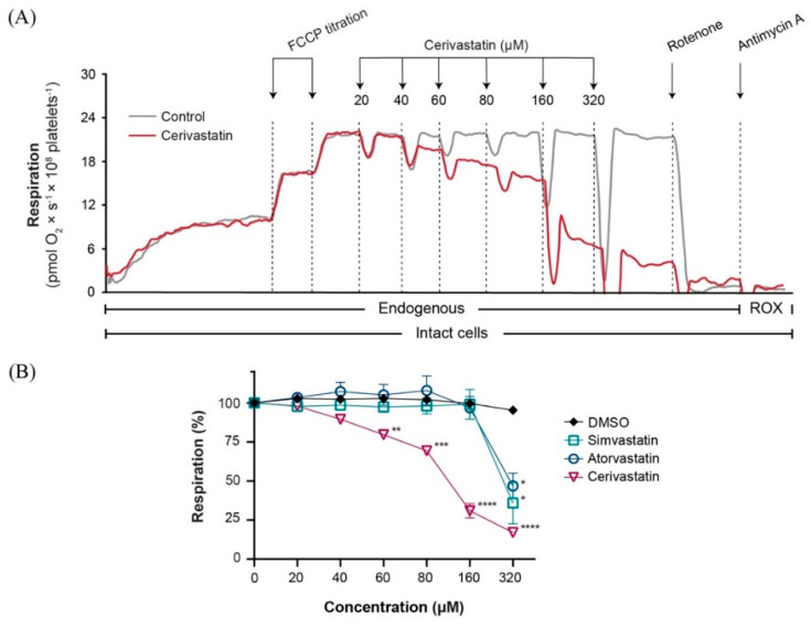
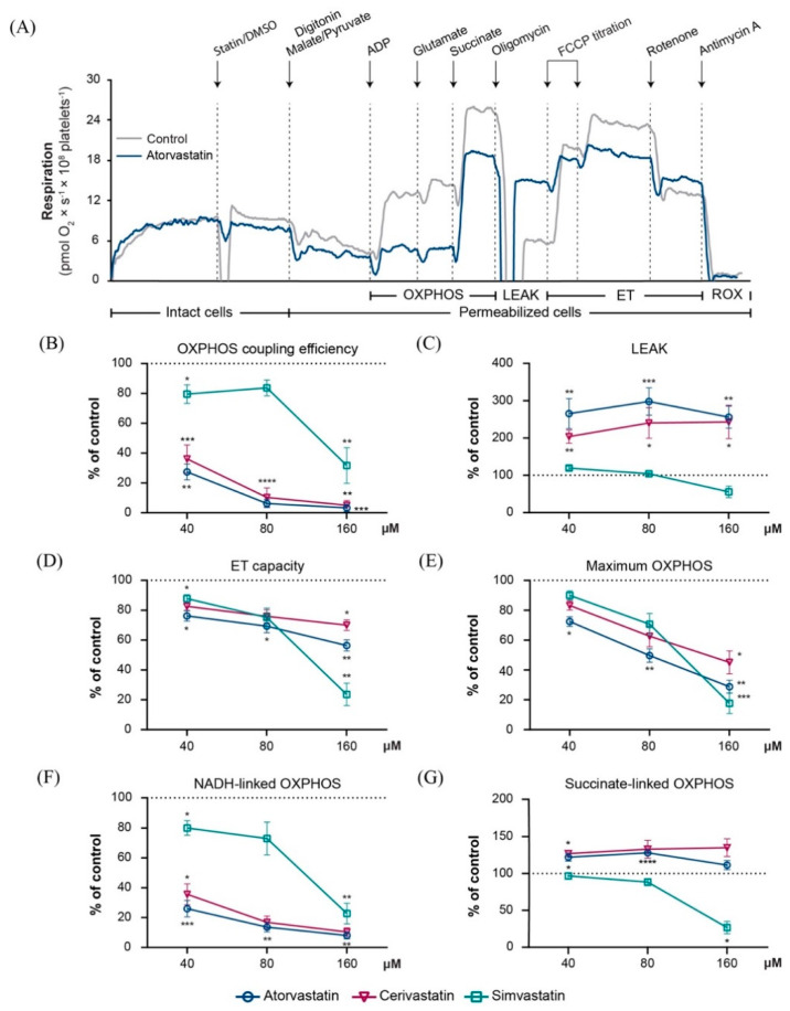
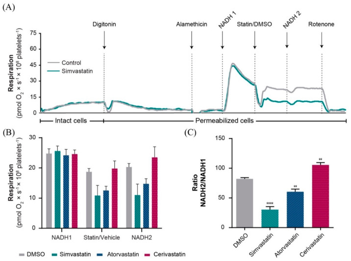
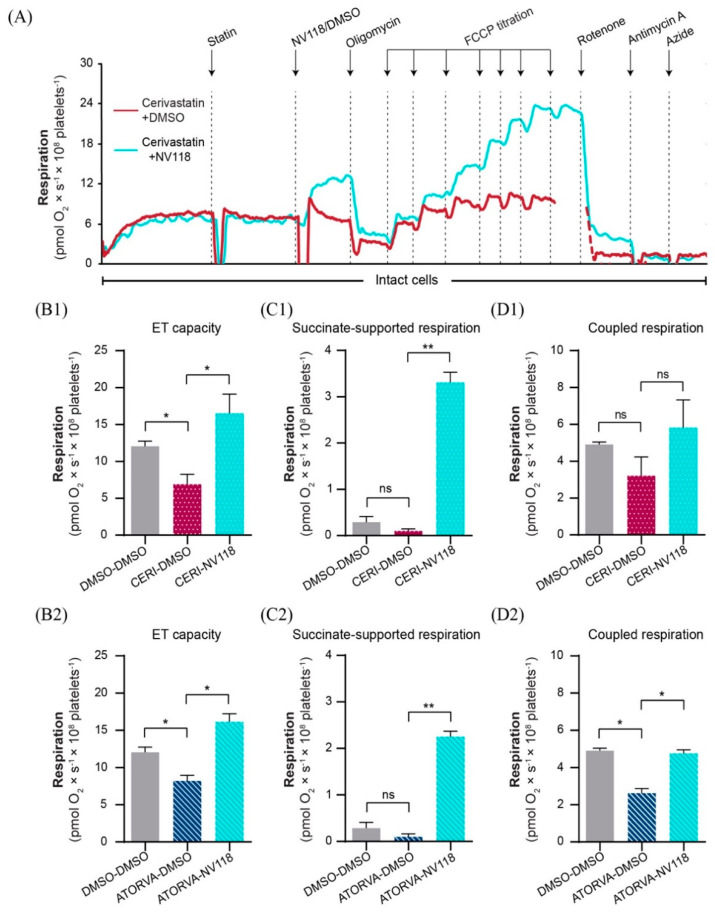
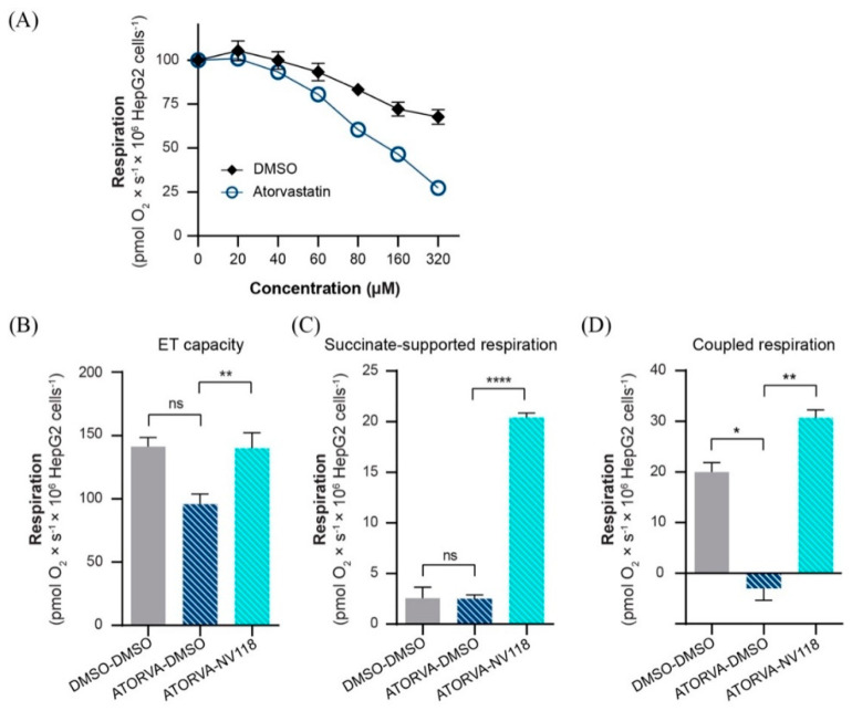
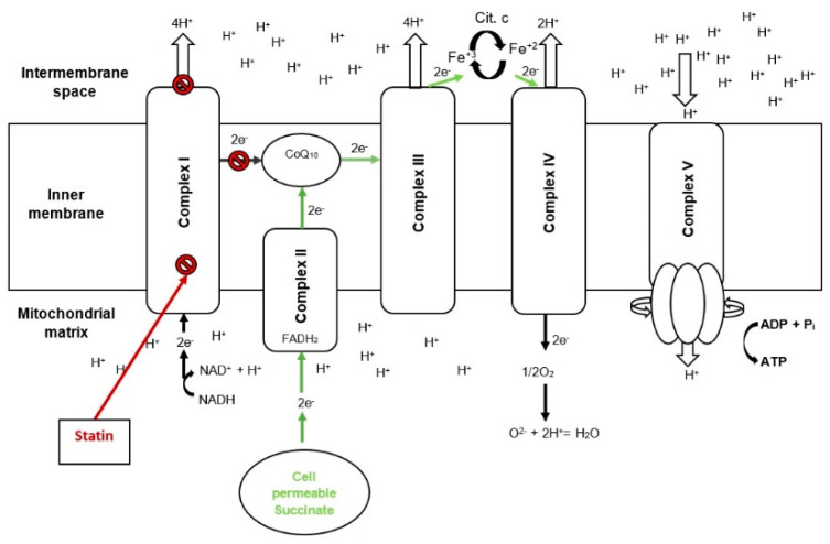
Statins are the cornerstone of lipid-lowering therapy. Although generally well tolerated, statin-associated muscle symptoms (SAMS) represent the main reason for treatment discontinuation. Mitochondrial dysfunction of complex I has been implicated in the pathophysiology of SAMS. The present study proposed to assess the concentration-dependent ex vivo effects of three statins on mitochondrial respiration in viable human platelets and to investigate whether a cell-permeable prodrug of succinate (complex II substrate) can compensate for statin-induced mitochondrial dysfunction. Mitochondrial respiration was assessed by high-resolution respirometry in human platelets, acutely exposed to statins in the presence/absence of the prodrug NV118. Statins concentration-dependently inhibited mitochondrial respiration in both intact and permeabilized cells. Further, statins caused an increase in non-ATP generating oxygen consumption (uncoupling), severely limiting the OXPHOS coupling efficiency, a measure of the ATP generating capacity. Cerivastatin (commercially withdrawn due to muscle toxicity) displayed a similar inhibitory capacity compared with the widely prescribed and tolerable atorvastatin, but did not elicit direct complex I inhibition. NV118 increased succinate-supported mitochondrial oxygen consumption in atorvastatin/cerivastatin-exposed platelets leading to normalization of coupled (ATP generating) respiration. The results acquired in isolated human platelets were validated in a limited set of experiments using atorvastatin in HepG2 cells, reinforcing the generalizability of the findings.

Keywords: HepG2 cells; NV118; cell-permeable succinate; mitochondria; platelets; statins.


Conflict of interest statement

I.C., E.Å.F., J.K.E., M.J.H. and E.E. have, or have had, salary from and/or equity interest in Abliva AB (previously named NeuroVive Pharmaceutical AB), a company active in the field of mitochondrial medicine. J.K.E., E.E., and M.J.H. have filed patent applications for the use of succinate prodrugs for treatment of lactic acidosis or drug-induced side effects due to complex I-related impairment of mitochondrial oxidative phosphorylation (WO/2015/155238) and protected carboxylic acid-based metabolites for treatment of mitochondrial disorders (WO/2017/060400, WO/2017/060418, WO/2017/060422). This does not alter our adherence to manuscript policies on sharing data and materials. Abliva AB had no role in the study design, the data collection and analysis, or the preparation of the manuscript. The remaining authors declare that they have no known competing financial interests or personal relationships that could have appeared to influence the work reported in this paper.

Figures

Figure 1
Figure 1
Statins induced a concentration−dependent inhibition of mitochondrial respiration in intact human platelets. (A) Representative traces of cerivastatin (red) and DMSO (grey) titration. Respiration of human platelets was measured after sub-maximal uncoupling with FCCP (2 µM). (B) The concentration-dependent effect of simvastatin (open square), atorvastatin (open circle) and cerivastatin (open triangle) was measured by titrating increasing concentrations of statin or vehicle (DMSO) (black rhombus). Data is expressed as mean±SEM. FCCP, carbonyl cyanide p-(trifluoromethoxy) phenylhydrazone; DMSO, dimethyl sulfoxide; ROX, residual oxygen consumption. Two-way ANOVA with Bonferroni post hoc test was performed on antimycin-corrected data. * p < 0.05; ** p < 0.01; *** p < 0.001; **** p < 0.0001 vs. DMSO.
Figure 2
Figure 2
Statins induced a concentration−dependent inhibition of mitochondrial respiration in permeabilized human platelets. (A) Representative traces of atorvastatin 40 µM (blue) and DMSO (grey). Concentration-dependent effects were assessed for 3 concentrations (40 µM, 80 µM and 160 µM, respectively) of atorvastatin (open circle), cerivastatin (open triangle) and simvastatin (open square), respectively. OXPHOS coupling efficiency (B), LEAK (C), maximal ET capacity (D), maximal OXPHOS (E), NADH-linked OXPHOS (F), and succinate-linked OXPHOS (G) capacities were evaluated. Data is expressed as mean±SEM of the percent of control (platelets exposed to the corresponding volume of DMSO for each of the 3 concentrations of statin). Two-way ANOVA with Bonferroni post hoc test was performed on antimycin-corrected data. DMSO, dimethyl sulfoxide; ET, electron transport; LEAK, non-phosphorylating resting stat; OXPHOS, oxidative phosphorylation; ROX, residual oxygen consumption. * p < 0.05; ** p < 0.01; *** p < 0.001; **** p < 0.0001 vs. DMSO.
Figure 3
Figure 3
Statin effects on NADH−linked oxygen consumption in alamethicin-permeabilized platelet mitochondria. (A) Representative trace of simvastatin 160 µM (green) and DMSO (grey). (B) Oxygen consumption of permeabilized platelet mitochondria was measured at three points: Figure 0. mM) next in the presence of the statin (160 µM)/DMSO control (DMSO-grey bar, simvastatin-green bar, atorvastatin-blue bar, cerivastatin-red bar) and then after the addition of statin plus a second dose of NADH (0.75 mM). (C) Calculated NADH-linked oxygen consumption (∆) for each statin and DMSO control, evaluated as the ratio between the two consecutive additions of NADH (NADH2/NADH1). Data is expressed as mean ± SEM of rotenone-corrected data, one-way ANOVA with Bonferroni post hoc test. DMSO, dimethyl sulfoxide. ** p < 0.01; **** p < 0.001 vs. DMSO.
Figure 4
Figure 4
The effects of NV118 on statin−dependent respiratory dysfunction in human platelets. (A) Representative overlay trace of statin-exposed platelets in the absence (red) or presence (blue) of the succinate prodrug NV118. NV118 effects in cerivastatin (B1,C1,D1) and atorvastatin (B2,C2,D2) exposed platelets were measured as compared to its vehicle (DMSO). As negative control of the experiment, platelets were exposed only to DMSO (DMSO-DMSO). Data is expressed as mean±SEM. One-way ANOVA with Bonferroni post hoc test was performed on antimycin-corrected data. ATORVA, atorvastatin; CERI, cerivastatin; DMSO, dimethyl sulfoxide; ET, electron transport. ns = no significance; * p < 0.05; ** p < 0.01.
Figure 5
Figure 5
The effects of NV118 on statin−dependent respiratory dysfunction in HepG2 cells. (A) Respiration was assessed after sub-maximal uncoupling with FCCP (4 µM) followed by increasing concentrations of atorvastatin or vehicle (DMSO). NV118 effects on atorvastatin exposed HepG2 cells (BD) were measured as compared to its vehicle (DMSO). As negative control of the experiment HepG2 cells were exposed only to DMSO (DMSO-DMSO). Data is expressed as mean ± SEM. One-way ANOVA with Bonferroni post hoc test was performed on antimycin-corrected data. ATORVA, atorvastatin; DMSO, dimethyl sulfoxide; ET, electron transport. ns = no significance; * p < 0.05; ** p < 0.01; **** p < 0.0001.
Figure 6
Figure 6
Cell permeable succinate bypasses statin−induced complex I mitochondrial dysfunction. Scheme 10. ubiquinone; e, electron; FADH2, dihydroflavine-adenine dinucleotide; Fe+3, iron(III); Fe+2, iron(II); H+, proton; NAD+, Nicotinamide adenine dinucleotide oxidized form; NADH, Nicotinamide adenine dinucleotide reduced form; OXPHOS, oxidative phosphorylation.

References

    1. Benjamin E.J., Muntner P., Alonso A., Bittencourt M.S., Callaway C.W., Carson A.P., Chamberlain A.M., Chang A.R., Cheng S., Das S.R., et al. Heart Disease and Stroke Statistics—2019 Update: A Report From the American Heart Association. Circulation. 2019;139:e56–e528. doi: 10.1161/CIR.0000000000000659. - DOI - PubMed
    1. Mach F., Baigent C., Catapano A.L., Koskinas K.C., Casula M., Badimon L., Chapman M.J., De Backer G.G., Delgado V., Ference B.A., et al. 2019 ESC/EAS Guidelines for the management of dyslipidaemias: Lipid modification to reduce cardiovascular risk: The Task Force for the management of dyslipidaemias of the European Society of Cardiology (ESC) and European Atherosclerosis Society (EAS) Eur. Heart J. 2019 doi: 10.1093/eurheartj/ehz455. - DOI - PubMed
    1. Ference B.A., Ginsberg H.N., Graham I., Ray K.K., Packard C.J., Bruckert E., Hegele R.A., Krauss R.M., Raal F.J., Schunkert H., et al. Low-density lipoproteins cause atherosclerotic cardiovascular disease. 1. Evidence from genetic, epidemiologic, and clinical studies. A consensus statement from the European Atherosclerosis Society Consensus Panel. Eur. Heart J. 2017;38:2459–2472. doi: 10.1093/eurheartj/ehx144. - DOI - PMC - PubMed
    1. Björkhem-Bergman L., Lindh J.D., Bergman P. What is a relevant statin concentration in cell experiments claiming pleiotropic effects? Br. J. Clin. Pharm. 2011;72:164–165. doi: 10.1111/j.1365-2125.2011.03907.x. - DOI - PMC - PubMed
    1. Stroes E.S., Thompson P.D., Corsini A., Vladutiu G.D., Raal F.J., Ray K.K., Roden M., Stein E., Tokgözoğlu L., Nordestgaard B.G., et al. Statin-associated muscle symptoms: Impact on statin therapy-European Atherosclerosis Society Consensus Panel Statement on Assessment, Aetiology and Management. Eur. Heart J. 2015;36:1012–1022. doi: 10.1093/eurheartj/ehv043. - DOI - PMC - PubMed

Substances

LinkOut - more resources