. 2021 Jan 2;14(1):181.
doi: 10.3390/ma14010181.

Design of 3D Additively Manufactured Hybrid Structures for Cranioplasty

Affiliations

Design of 3D Additively Manufactured Hybrid Structures for Cranioplasty

Roberto De Santis et al. Materials (Basel). .

Abstract

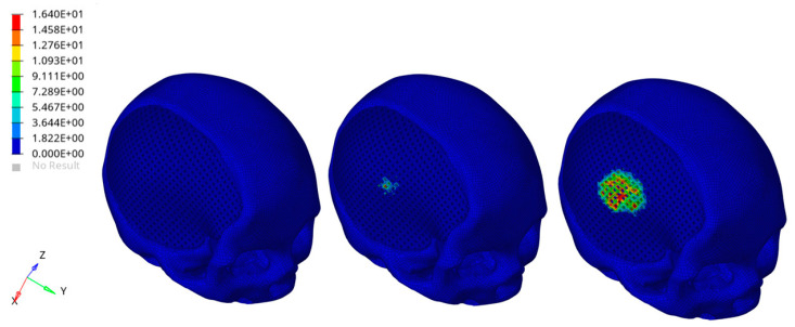
A wide range of materials has been considered to repair cranial defects. In the field of cranioplasty, poly(methyl methacrylate) (PMMA)-based bone cements and modifications through the inclusion of copper doped tricalcium phosphate (Cu-TCP) particles have been already investigated. On the other hand, aliphatic polyesters such as poly(ε-caprolactone) (PCL) and polylactic acid (PLA) have been frequently investigated to make scaffolds for cranial bone regeneration. Accordingly, the aim of the current research was to design and fabricate customized hybrid devices for the repair of large cranial defects integrating the reverse engineering approach with additive manufacturing, The hybrid device consisted of a 3D additive manufactured polyester porous structures infiltrated with PMMA/Cu-TCP (97.5/2.5 w/w) bone cement. Temperature profiles were first evaluated for 3D hybrid devices (PCL/PMMA, PLA/PMMA, PCL/PMMA/Cu-TCP and PLA/PMMA/Cu-TCP). Peak temperatures recorded for hybrid PCL/PMMA and PCL/PMMA/Cu-TCP were significantly lower than those found for the PLA-based ones. Virtual and physical models of customized devices for large cranial defect were developed to assess the feasibility of the proposed technical solutions. A theoretical analysis was preliminarily performed on the entire head model trying to simulate severe impact conditions for people with the customized hybrid device (PCL/PMMA/Cu-TCP) (i.e., a rigid sphere impacting the implant region of the head). Results from finite element analysis (FEA) provided information on the different components of the model.

Keywords: composite bone cement for cranioplasty; design for additive manufacturing; finite element analysis; reverse engineering; temperature profile analysis.


Conflict of interest statement

The authors declare no conflict of interest.

Figures

Figure 1
Figure 1
Typical image of a 3D printed porous structure (diameter of 10 mm, height of about 5 mm) (left); 3D porous structure incorporating a k-type thermocouple (center); hollow cylindrical Teflon mold equipped with k-type thermocouple (right).
Figure 2
Figure 2
Three-dimensional reconstruction of a skull with a large cranial defect. The images were analyzed starting from a previous 3D scanning process [1].
Figure 3
Figure 3
Three-dimensional geometrical models of a customized device for large cranial defect: non-porous model (left); model with interconnected pore network (right).
Figure 4
Figure 4
Three-dimensional additive manufactured devices for large cranial defect: non-porous model (left); model with interconnected pore network (right).
Figure 5
Figure 5
Three-dimensional geometrical models of a customized hybrid device for large cranial defect: top view evidencing the external cement layer (left); bottom view highlighting the cement infiltration in the fully interconnected pore network (right). Green color was chosen to identify the cement component of the hybrid device.
Figure 6
Figure 6
A simplified image reporting some components of the finite element analysis (FEA) model.
Figure 7
Figure 7
Temperature peaks measured during setting in the case of plain bone cements (PMMA, PMMA/Cu-TCP) and hybrid structures consisting of 3D polylactic acid (PLA) or poly(ε-caprolactone) (PCL) networks infiltrated with cement. Statistical analysis was performed by analysis of variance (ANOVA). Statistical significance was set at p < 0.05.
Figure 8
Figure 8
An example of temperature profiles: plain bone cement, PLA and PCL structures infiltrated with PMMA, evidencing some differences during the cooling phase.
Figure 9
Figure 9
Images of the 3D physical model of a skull with a large cranial defect, which was previously fabricated by inkjet printing, starting from image capture and analysis techniques [1]: top-front view (left) and lateral view (right).
Figure 10
Figure 10
Feasibility of the reported technical solutions: images of virtual (left) and physical (center) models of skull with 3D additive manufactured PCL porous structure for large cranial defect 3D porous structure; image of virtual model of skull with 3D hybrid device (3D PCL porous structure infiltrated with PMMA/Cu-TCP 97.5/2.5 bone cement) for large cranial defect (right).
Figure 11
Figure 11
Biomechanical response as a result of the rigid sphere impacting the implant region of the head: von Mises stress (MPa) distributions for the external cement layer of the hybrid device at three different times after the initial impact (left). The figure is a guide for the eye to see the effect of the impact for the selected component. The color scale was chosen to allow for comparison among the models ad different times.
Figure 12
Figure 12
Biomechanical response as a result of the rigid sphere impacting the implant zone region of the head: von Mises stress (MPa) distributions for the 3D PCL structure underneath the external cement layer and infiltrated with the bone cement, at three different times after the initial impact (left). Bone cement was removed to visualize the effect of the impact for the selected component. The color scale was chosen to allow for comparison among the models at different times.

References

    1. De Santis R., Gloria A., Ambrosio L. Materials and technologies for craniofacial tissue repair and regeneration. Top. Med. 2010;16:1–6.
    1. Piitulainen J.M., Kauko T., Aitasalo K.M., Vuorinen V., Vallittu P.K., Posti J.P. Outcomes of Cranioplasty with Synthetic Materials and Autologous Bone Grafts. World Neurosurg. 2015;83:708–714. doi: 10.1016/j.wneu.2015.01.014. - DOI - PubMed
    1. Unterhofer C., Wipplinger C., Verius M., Recheis W., Thomé C., Ortler M. Reconstruction of large cranial defects with poly-methyl-methacrylate (PMMA) using a rapid prototyping model and a new technique for intraoperative implant modeling. Neurol. Neurochir. Polska. 2017;51:214–220. doi: 10.1016/j.pjnns.2017.02.007. - DOI - PubMed
    1. Lee S.C., Wu C.T., Lee S.T., Chen P.J. Cranioplasty using polymethyl methacrylate prostheses. J. Clin. Neurosci. 2009;16:56–63. doi: 10.1016/j.jocn.2008.04.001. - DOI - PubMed
    1. Harris D.A., Fong A.J., Buchanan E.P., Monson L., Khechoyan D., Lam S. History of synthetic materials in alloplastic cranioplasty. Neurosurg. Focus. 2014;36:E20. doi: 10.3171/2014.2.FOCUS13560. - DOI - PubMed

LinkOut - more resources