

Scanning and Actuation Techniques for Cantilever-Based Fiber Optic Endoscopic Scanners-A Review
- PMID: 33401728
- PMCID: PMC7795415
- DOI: 10.3390/s21010251
Scanning and Actuation Techniques for Cantilever-Based Fiber Optic Endoscopic Scanners-A Review
Abstract
Endoscopes are used routinely in modern medicine for in-vivo imaging of luminal organs. Technical advances in the micro-electro-mechanical system (MEMS) and optical fields have enabled the further miniaturization of endoscopes, resulting in the ability to image previously inaccessible small-caliber luminal organs, enabling the early detection of lesions and other abnormalities in these tissues. The development of scanning fiber endoscopes supports the fabrication of small cantilever-based imaging devices without compromising the image resolution. The size of an endoscope is highly dependent on the actuation and scanning method used to illuminate the target image area. Different actuation methods used in the design of small-sized cantilever-based endoscopes are reviewed in this paper along with their working principles, advantages and disadvantages, generated scanning patterns, and applications.
Keywords: MEMS actuators; electromagnetic; electrostatic; electrothermal; endoscopes; medical imaging; piezoelectric; scanning patterns; shape memory alloys.
Conflict of interest statement
The authors declare no conflict of interest.
Figures
Similar articles
-
Submillimeter Sized 2D Electrothermal Optical Fiber Scanner.Sensors (Basel). 2022 Dec 30;23(1):404. doi: 10.3390/s23010404. Sensors (Basel). 2022. PMID: 36617001 Free PMC article.
-
Real-time Lissajous imaging with a low-voltage 2-axis MEMS scanner based on electrothermal actuation.Opt Express. 2020 Mar 16;28(6):8512-8527. doi: 10.1364/OE.380690. Opt Express. 2020. PMID: 32225475
-
Scanning Micromirror Platform Based on MEMS Technology for Medical Application.Micromachines (Basel). 2016 Feb 6;7(2):24. doi: 10.3390/mi7020024. Micromachines (Basel). 2016. PMID: 30407397 Free PMC article. Review.
-
Ultralow-voltage electrothermal MEMS based fiber-optic scanning probe for forward-viewing endoscopic OCT.Opt Lett. 2019 May 1;44(9):2232-2235. doi: 10.1364/OL.44.002232. Opt Lett. 2019. PMID: 31042191 Free PMC article.
-
A Review of Actuation and Sensing Mechanisms in MEMS-Based Sensor Devices.Nanoscale Res Lett. 2021 Jan 26;16(1):16. doi: 10.1186/s11671-021-03481-7. Nanoscale Res Lett. 2021. PMID: 33496852 Free PMC article. Review.
Cited by
-
Design and test of a rigid endomicroscopic system for multimodal imaging and femtosecond laser ablation.J Biomed Opt. 2023 Jun;28(6):066004. doi: 10.1117/1.JBO.28.6.066004. Epub 2023 Jun 28. J Biomed Opt. 2023. PMID: 37388219 Free PMC article.
-
Structural Design and Experimental Studies of Resonant Fiber Optic Scanner Driven by Co-Fired Multilayer Piezoelectric Ceramics.Micromachines (Basel). 2023 Feb 23;14(3):517. doi: 10.3390/mi14030517. Micromachines (Basel). 2023. PMID: 36984924 Free PMC article.
-
Passively scanned, single-fiber optical coherence tomography probes for gastrointestinal devices.Lasers Surg Med. 2022 Sep;54(7):935-944. doi: 10.1002/lsm.23576. Epub 2022 Jun 16. Lasers Surg Med. 2022. PMID: 35708124 Free PMC article.
-
Towards OCT-Guided Endoscopic Laser Surgery-A Review.Diagnostics (Basel). 2023 Feb 11;13(4):677. doi: 10.3390/diagnostics13040677. Diagnostics (Basel). 2023. PMID: 36832167 Free PMC article. Review.
-
Wide-field endoscope accessory for multiplexed fluorescence imaging.Sci Rep. 2023 Nov 9;13(1):19527. doi: 10.1038/s41598-023-45955-x. Sci Rep. 2023. PMID: 37945660 Free PMC article.
References
-
- WHO, Cancer, World Health Organization, 12 09 2018. [(accessed on 6 October 2020)]; Available online: https://www.who.int/news-room/fact-sheets/detail/cancer.
-
- Stewart B.W., Wild C.P. World Cancer Report 2014. IARC; Lyon, France: 2014.
-
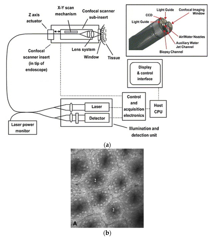
- Conigliaro R., Pigò F. New Techniques in Gastrointestinal Endoscopy. IntechOpen; London, UK: 2011. New Techniques in Endoscopy: Confocal Laser Endomicroscopy; pp. 213–230.
Publication types
LinkOut - more resources
Full Text Sources
Other Literature Sources
