

doi: 10.14245/ns.2040556.278.
Epub 2020 Dec 31.
The Art of Diagnosis in the Cervical Spine
Affiliations
- PMID: 33401849
- PMCID: PMC7788414
- DOI: 10.14245/ns.2040556.278
Item in Clipboard
The Art of Diagnosis in the Cervical Spine
Neurospine.
2020 Dec.
No abstract available
Conflict of interest statement
The authors have nothing to disclose.
Figures
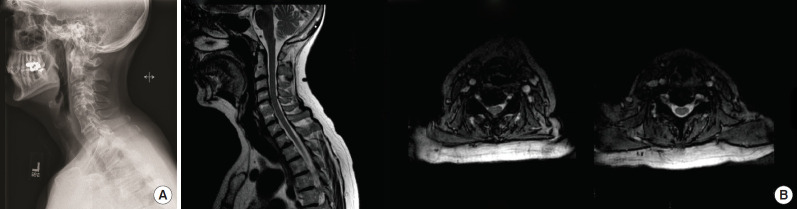
(A) Sagittal radiograph of the cervical spine demonstrating a fusion of C5–6 with adjacent segment degenerative disease at C4–5. (B) Left image: sagittal T2-weighted image of the cervical spine. Center image: axial T2-weighted imaging of the C4–5 level demonstrating right-sided foraminal disc herniation at the C4–5 level. Right image: axial T2-weighted imaging of the fused C5–6 level does not demonstrate any significant disc pathology to explain the patient’s left-sided symptoms.
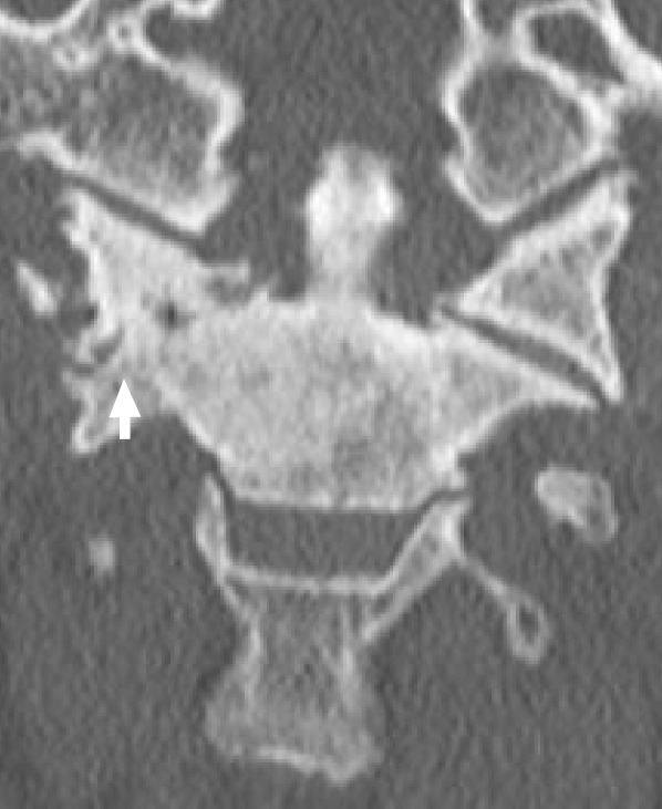
Coronal computed tomography imaging demonstrating C1–2 arthritis on patient’s right side (withe arrow).
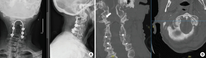
Imaging from a patient presented after multiple cervical surgeries with persistent high cervical pain at the base of the occiput on the right side. (A) X-rays show prior instrumentation in place (anteroposterior view on left, lateral view on right). (B) Computer tomography images (coronal reformat on left, axial reformat on right) show severe right atlanto-occipital arthritis (white arrow). Pain resolved after right atlanto-occipital steroid injections.
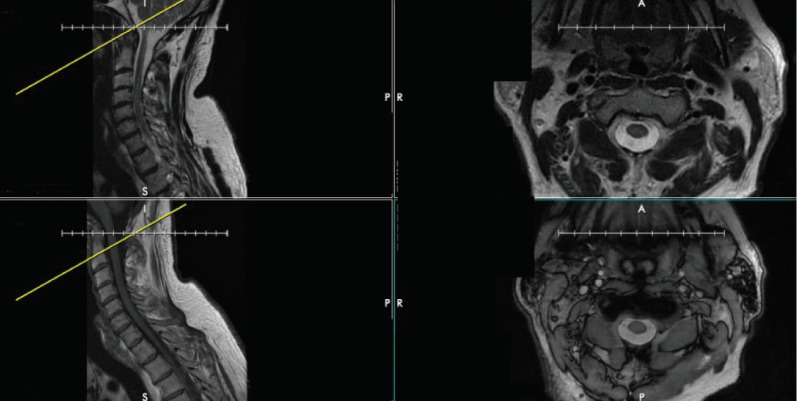
The discrepancy between axial and sagittal cuts on magnetic resonance imaging of the cervical spine. Top left and bottom left show a T2- and T1-weighted sagittal cuts, respectively, demonstrating 2 different cervical spine levels. However, the top right and bottom right images show T2- and T1-weighted axial cuts, respectively, that both are at the same level (at the base of C2).
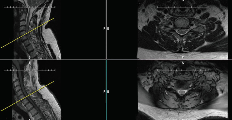
The discrepancy between reference cuts on different MRI sequences of the cervical spine. The top and bottom right images show T2- and T1-weighted axial cuts at the same level in the cervical spine, but the reference lines on the sagittal T2- and T1-weighted images seen on the top left and bottom left images differ by one level entirely, with the T2-weighted image showing the C5-6 level and the T1-weighted image showing the C6-7 level.
References
-
- Schwartz HG. Anastomoses between cervical nerve roots. J Neurosurg. 1956;13:190–4. - PubMed
-
- Nittby HR, Bendix T. On the variations of cervical dermatomes. Int J Anat Res. 2014:462–9.
-
- Kimura J. Electrodiagnosis in diseases of nerve and muscle: principles and practice. 3rd ed. New York: Oxford University Press; 2001.
-
- Dumitru D, Amato A, Zwarts M. Electrodiagnostic medicine. 2nd ed. Philadelphia (PA): Hanley and Belfus; 2002.
-
- Greathouse DG, Joshi A. Radiculopathy of the eighth cervical nerve. J Orthop Sports Phys Ther. 2010;40:811–7. - PubMed
LinkOut - more resources
Full Text Sources
