. 2021 Jan 5;14(664):eabc8521.
doi: 10.1126/scisignal.abc8521.

The Campylobacter jejuni chemoreceptor Tlp10 has a bimodal ligand-binding domain and specificity for multiple classes of chemoeffectors

Affiliations

The Campylobacter jejuni chemoreceptor Tlp10 has a bimodal ligand-binding domain and specificity for multiple classes of chemoeffectors

Bassam A Elgamoudi et al. Sci Signal. .

Abstract

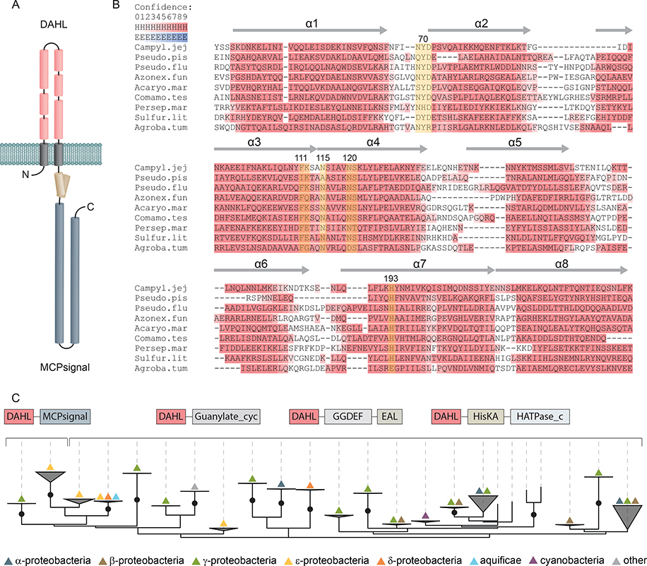
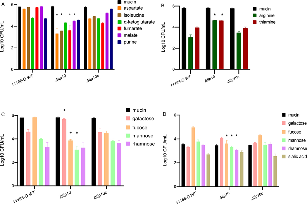
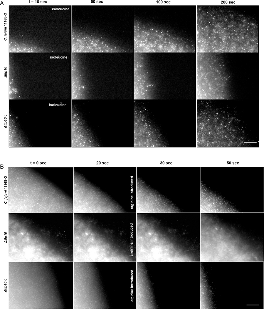
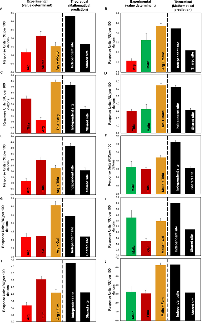
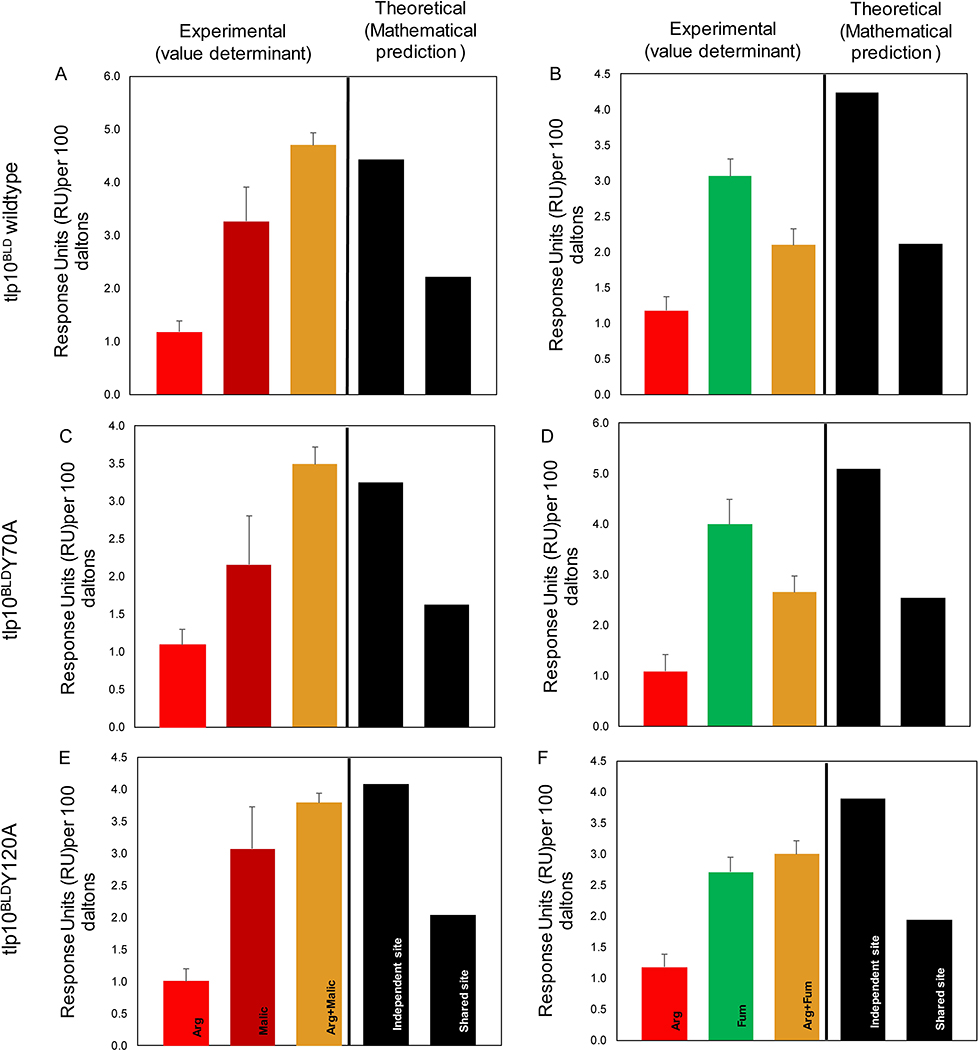
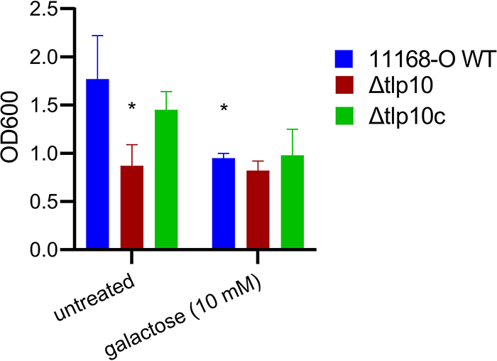
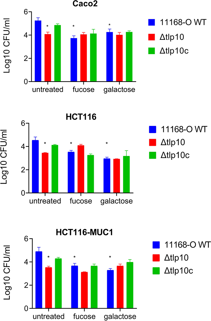
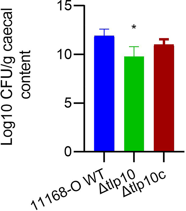
Campylobacter jejuni is a bacterial pathogen that is a common cause of enteritis in humans. We identified a previously uncharacterized type of sensory domain in the periplasmic region of the C. jejuni chemoreceptor Tlp10, termed the DAHL domain, that is predicted to have a bimodular helical architecture. Through two independent ligand-binding sites in this domain, Tlp10 responded to molecular aspartate, isoleucine, fumarate, malate, fucose, and mannose as attractants and to arginine, galactose, and thiamine as repellents. Tlp10 also recognized glycan ligands when present as terminal and intermediate residues of complex structures, such as the fucosylated human ganglioside GM1 and Lewisa antigen. A tlp10 mutant strain lacking the ligand-binding sites was attenuated in its ability to colonize avian caeca and to adhere to cultured human intestinal cells, indicating the potential involvement of the DAHL domain in host colonization and disease. The Tlp10 intracellular signaling domain interacted with the scaffolding proteins CheV and CheW, which couple chemoreceptors to intracellular signaling machinery, and with the signaling domains of other chemoreceptors, suggesting a key role for Tlp10 in signal transduction and incorporation into sensory arrays. We identified the DAHL domain in other bacterial signal transduction proteins, including the essential virulence induction protein VirA from the plant pathogen Agrobacterium tumefaciens Together, these results suggest a potential link between Tlp10 and C. jejuni virulence.


Conflict of interest statement

Competing interests: The authors declare that they have no competing interests.

Figures

Fig. 1.
Fig. 1.. Secondary structure prediction and phyletic distribution of the DAHL domain.
(A) Schematic representation of Tlp10 topology with the predicted architecture of the DAHL domain as the LBD, the transmembrane region, and the intracellular MCP signaling domain. (B) MSA and secondary structure prediction of periplasmic DAHL domains from various bacterial species. Predicted α-helical regions are labelled and highlighted in red. Conserved amino acid positions are highlighted in yellow. Representative sequences were selected from different clades on the phylogenetic tree. Campyl.jej: C. jejuni, Tlp10 WP_002865311.1; Pseudo.pis: Pseudoalteromonas piscicida, WP_099642651.1; Pseudo.flu: Pseudomonas fluorescens, WP_003172050.1; Azonex.fun: Azonexus fungiphilus, WP_121456889.1; Acaryo.mar: Acaryochloris marina, WP_012165516.1; Comamo.tes: Comamonas testosterone, WP_003076421.1; Persep.mar: Persephonella marina, WP_012676181.1; Sulfur.lit: Sulfurovum lithotrophicum, WP_052746078.1; Agroba.tum: A. tumefaciens, VirA WP_012478065.1. (C) Maximum likelihood phyletic distribution and protein architecture variations of DAHL domain–containing proteins with bootstrap support based on 500 replicates. Neighboring domains are indicated according to Pfam database nomenclature: MCPsignal, MCP signaling domain, PF00015; Guanylate_cyc, adenylate and guanylate cyclase catalytic domain, PF00211; GGDEF, diguanylate cyclase, PF00990; EAL, diguanylate phosphodiesterase, PF00563; HisKA, histidine kinaseA (phospho-acceptor) domain, PF00512; HATPase_c; -, DNA gyrase B-, phytochrome-like ATPases, PF02518. Bootstrap values more than 70% are indicated by black circles.
Fig. 2.
Fig. 2.. Chemotaxis assays with wild-type and Tlp10 mutant C. jejuni.
(A and B) Wild-type C. jejuni strain 11168-O (11168-O WT), the isogenic Tlp10LBD mutant (Δtlp10), and the complemented mutant (Δtlp10c) were subjected to chemotaxis assays measuring the movement of starved cells towards test ligands considered attractants (A) and ligands considered repellents (B). (C) Chemotaxis to the monosaccharide ligands galactose, fucose, mannose, and rhamnose. The universal attractant mucin was used a positive control. The C. jejuni non-motile mutant (81116ΔflaA/flaB) and PBS were used as negative controls (Table S5). (D) μ-slide chemotaxis assays testing motility within a gradient of the test ligand were performed using galactose, fucose, mannose, rhamnose, and sialic acid as the test compounds. All data (Log 10 CFU/mL) represent three independent experiments, each performed in triplicate (n = 3). Asterisks indicate significant differences (P < 0.05; t-test).
Fig. 3.
Fig. 3.. Migration of 11168-O WT, Δtlp10, and Δtlp10c in response to chemoeffectors.
Time-lapse imaging of fluorescently labelled C. jejuni 11168-O (WT), Δtlp10, and Δtlp10c cells migrating from the left chamber toward the right chamber along a chemoeffector gradient on μ-Slide Chemotaxis chamber. (A) Attractant response toward 10 mM isoleucine. (B) Response of cells that had partially migrated toward 10 mM isoleucine before the introduction of 10 mM arginine into the right chamber at 20 sec. This figure is representative of five independent experiments (n=5). Scale bars, 100 μm.
Fig. 4.
Fig. 4.. Competition SPR analysis of Tlp10LBD.
Competition analysis of the binding of arginine (Arg), malate (Malic), thiamine (Thia), galactose (Gal), fumarate (Fum), and α-ketoglutarate (Aketo) to WT Tlp10LBD immobilized on a sensor chip. (A) Responses to Arg only, Aketo only, and Arg after saturation with α-ketoglutarate. (B) Responses to Arg only, Malic only, and Arg following saturation with malate. (C) Responses to Thia only, Arg only, and Thia following saturation with arginine. (D) Responses to Thia only, Malic only, and Thia following saturation with malate. (E) Responses to Arg only, Thia only, and Arg following saturation with thiamine. (F) Responses to Malic only, Thia only, and Malic following saturation with Thia. (G) Responses to Arg only, Gal only, and Arg following saturation with Gal. (H) Responses to Malic only, Gal only, and Malic following saturation with Gal. (I) Responses to Arg only, Fum only, and Arg following saturation with Fum. (J) Responses to Malic only, Fum only, and Malic following saturation with Fum. Results are presented for analyte A alone and analyte B alone (red, dark red, and green), and competition assay between analytes A and B (gold). The theoretical values for independent binding of the two analytes to separate sites and for binding to a shared site are shown as response units (RU) values based on mathematical theory (black). All response data were normalized to 100 Da molecular weight for each analyte allowing direct comparison of responses.
Fig. 5.
Fig. 5.. Effect of point mutations of Tlp10LBD on ligand binding.
SPR competition analysis of the binding of arginine (Arg), malate (Malic), and fumarate (Fum) to immobilized WT and mutant Tlp10LBD proteins. (A) Responses of Tlp10LBD to Arg only, Malic only, and Arg following saturation with Malic. (B) Responses of Tlp10LBD to Arg only, Fum only, and Arg following saturation with Fum. (C) Responses of Tlp10LBDY70A to Arg only, Malic only, and Arg following saturation with Malic. (D) Responses of Tlp10LBDY70A to Arg only, Fum only, and Arg following saturation with Fum. (E) Responses of Tlp10LBDN120A to Arg only, Malic only, and Arg following saturation with Malic. (F) Responses of Tlp10LBDN120A to Arg only, Fum only, and Arg following saturation with Fum. Results are presented for analyte A alone and analyte B alone (red, dark red, and green), and competition assay between analytes A and B (gold). The theoretical values for independent binding of the two analytes to separate sites and for binding to a shared site are shown as response units (RU) values based on mathematical theory (black). All response data were normalized to 100 Da molecular weight for each analyte allowing direct comparison of responses.
Fig. 6.
Fig. 6.. Reduced biofilm formation of Δtlp10 C. jejuni.
Quantification of biofilm formation by wild-type C. jejuni strain 11168-O (11168-O WT), the isogenic Tlp10LBD mutant (Δtlp10), and Δtlp10 complemented \ with full-length Tlp10 (Δtlp10c), with and without 10 mM galactose. The data represents three independent experiments, each performed in triplicate (n = 3). Asterisks indicate significant differences (P < 0.05; t-test).
Fig. 7.
Fig. 7.. Reduced adhesion of Δtlp10 C. jejuni to mammalian cells.
Quantification of the adhesion of WT 11168-O C. jejuni (11168-O WT), the isogenic Tlp10LBD mutant (Δtlp10), and Δtlp10 complemented with full-length Tlp10 (Δtlp10c) to Caco2 cells, HCT116 cells, and HCT116 cells overexpressing MUC1 in the presence and absence of 5 mM of fucose or galactose. Adhesion analyses are presented as the mean of adhesion from three independent experiments, each performed in triplicate (n = 3). Asterisks indicate significant differences (P < 0.05; t-test).
Fig 8.
Fig 8.. Reduced colonization of chickens by Δtlp10 C. jejuni.
Chicks were orally infected with 108 CFU of WT C. jejuni (11168-O WT), the Tlp10LBD mutant (Δtlp10), or the complemented mutant (Δtlp10c) at day of hatch. Ceca were recovered at 7 days post-inoculation, homogenized, serially diluted, and plated on selective medium for C. jejuni enumeration. Counts are shown as log CFU/g of caecal contents. n = 7 chicks for each C. jejuni strain. Asterisks indicate significant differences (P < 0.05; t-test).

References

    1. Wadhams GH, Armitage JP, Making sense of it all: Bacterial chemotaxis. Nat Rev Mol Cell Biology 5, 1024–1037 (2004). - PubMed
    1. Lux R, Shi W, Chemotaxis-guided movements in bacteria. Crit. Rev. Oral Biol. Med. 15, 207–220 (2004). - PubMed
    1. Ketley JM, Pathogenesis of enteric infection by Campylobacter. Microbiology 143, 5–21 (1997). - PubMed
    1. Poly F, Guerry P, Pathogenesis of Campylobacter. Current opinion in gastroenterology 24, 27–31 (2008). - PubMed
    1. W. H. Organization, The global view of campylobacteriosis: report of an expert consultation, Utrecht, Netherlands. World Health Organization, (2012).

Publication types

MeSH terms

LinkOut - more resources