. 2021 Feb 10;41(6):1218-1241.
doi: 10.1523/JNEUROSCI.2167-20.2020. Epub 2021 Jan 5.

Neuron-Specific FMRP Roles in Experience-Dependent Remodeling of Olfactory Brain Innervation during an Early-Life Critical Period

Affiliations

Neuron-Specific FMRP Roles in Experience-Dependent Remodeling of Olfactory Brain Innervation during an Early-Life Critical Period

Randall M Golovin et al. J Neurosci. .

Abstract

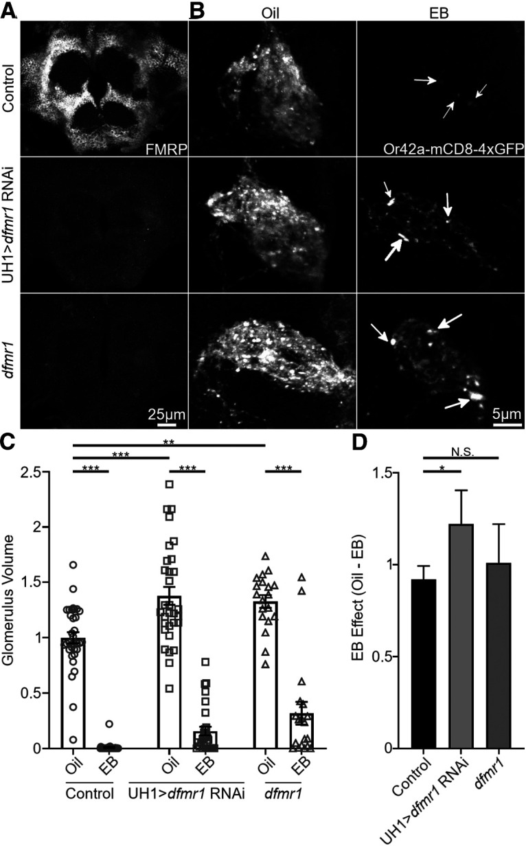
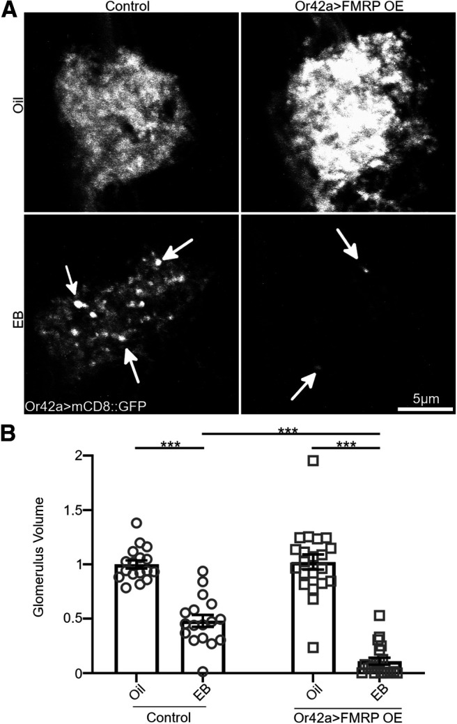
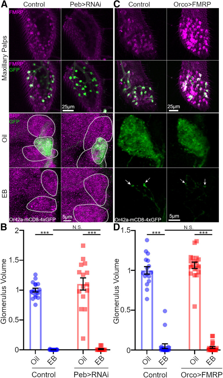
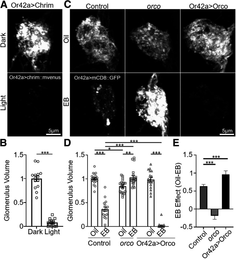
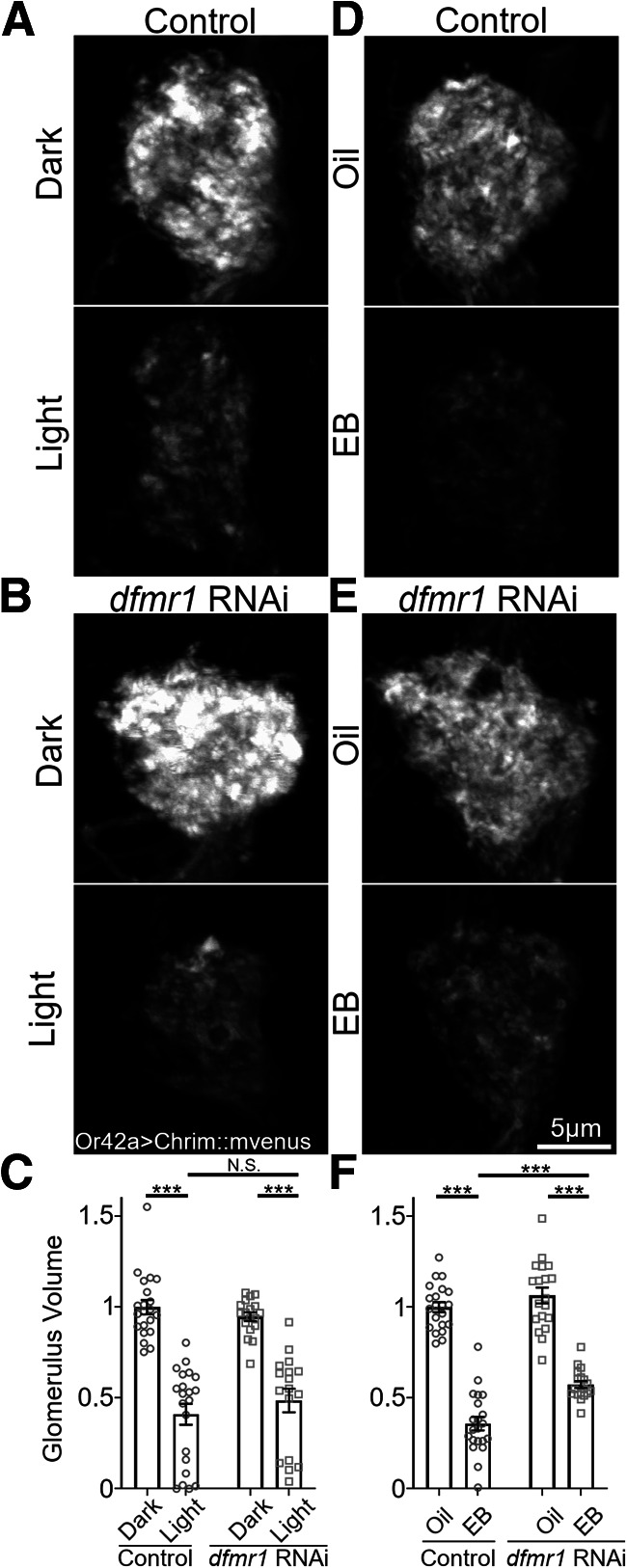
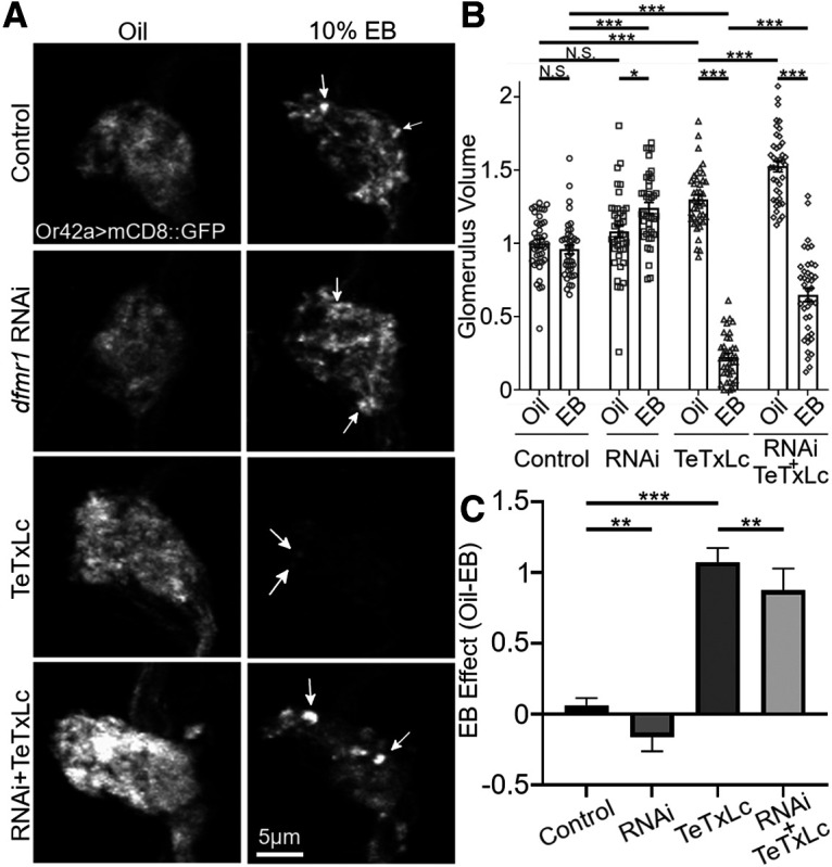
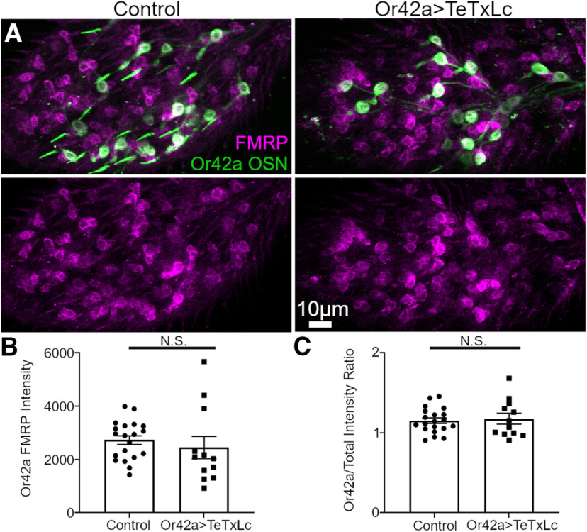
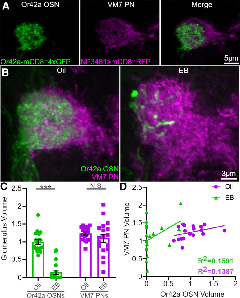
Critical periods are developmental windows during which neural circuits effectively adapt to the new sensory environment. Animal models of fragile X syndrome (FXS), a common monogenic autism spectrum disorder (ASD), exhibit profound impairments of sensory experience-driven critical periods. However, it is not known whether the causative fragile X mental retardation protein (FMRP) acts uniformly across neurons, or instead manifests neuron-specific functions. Here, we use the genetically-tractable Drosophila brain antennal lobe (AL) olfactory circuit of both sexes to investigate neuron-specific FMRP roles in the odorant experience-dependent remodeling of the olfactory sensory neuron (OSN) innervation during an early-life critical period. We find targeted OSN class-specific FMRP RNAi impairs innervation remodeling within AL synaptic glomeruli, whereas global dfmr1 null mutants display relatively normal odorant-driven refinement. We find both OSN cell autonomous and cell non-autonomous FMRP functions mediate odorant experience-dependent remodeling, with AL circuit FMRP imbalance causing defects in overall glomerulus innervation refinement. We find OSN class-specific FMRP levels bidirectionally regulate critical period remodeling, with odorant experience selectively controlling OSN synaptic terminals in AL glomeruli. We find OSN class-specific FMRP loss impairs critical period remodeling by disrupting responses to lateral modulation from other odorant-responsive OSNs mediating overall AL gain control. We find that silencing glutamatergic AL interneurons reduces OSN remodeling, while conversely, interfering with the OSN class-specific GABAA signaling enhances remodeling. These findings reveal control of OSN synaptic remodeling by FMRP with neuron-specific circuit functions, and indicate how neural circuitry can compensate for global FMRP loss to reinstate normal critical period brain circuit remodeling.SIGNIFICANCE STATEMENT Fragile X syndrome (FXS), the leading monogenic cause of intellectual disability and autism spectrum disorder (ASD), manifests severe neurodevelopmental delays. Likewise, FXS disease models display disrupted neurodevelopmental critical periods. In the well-mapped Drosophila olfactory circuit model, perturbing the causative fragile X mental retardation protein (FMRP) within a single olfactory sensory neuron (OSN) class impairs odorant-dependent remodeling during an early-life critical period. Importantly, this impairment requires activation of other OSNs, and the olfactory circuit can compensate when FMRP is removed from all OSNs. Understanding the neuron-specific FMRP requirements within a developing neural circuit, as well as the FMRP loss compensation mechanisms, should help us engineer FXS treatments. This work suggests FXS treatments could use homeostatic mechanisms to alleviate circuit-level deficits.

Keywords: Drosophila; critical period; fragile X mental retardation protein; fragile X syndrome; sensory experience; synapse elimination.


Figures

Figure 1.
Figure 1.
Maxillary palp (MP) to antennal lobe (AL) olfactory circuitry and neuron class-specific drivers A, Whole Drosophila head showing Or42a olfactory sensory neuron (OSN) innervation pattern (Or42a-Gal4>UAS-GtACR1-eYFP; green). OSN cell bodies in the maxillary palp (MP) project to the antennal lobe (AL). B, Or42a OSN innervation (Or42a-Gal4>UAS-CsChrimson::mVenus; green) of central brain (larger box in A) co-labeled for presynaptic Bruchpilot (BRP; magenta). Or42a OSNs extend axons via the labial nerve (bottom arrow) to terminate in the VM7 glomeruli (top arrow) of each AL. AL glomeruli postsynaptic projection neurons (PNs) send axons to the mushroom body (MB) and lateral horn (LH) in each brain hemisphere. C, Or42a OSN cell bodies (Or42a-mCD8-4xGFP; green) in MP (smaller box in (A) co-labeled for all OSN somata (Pebbled (Peb)-Gal4>UAS-mCD8::RFP; magenta). D, Or42a OSN axonal termini (Or42a-mCD8-4xGFP; green) and postsynaptic PNs (NP3481-Gal4>UAS-mCD8::RFP; magenta) in the AL VM7 (white box in (B). E, Neuron class-specific Gal4 drivers expressing UAS-mCD8::GFP in presynaptic OSNs (Peb-Gal4; top left), postsynaptic PNs (GH146-Gal4; top right), GABAergic local interneurons (LNs, NP1227-Gal4; bottom left) and Glutamatergic LNs (OK107-Gal4; bottom right) of the brain AL.
Figure 2.
Figure 2.
Or42a OSN-specific FMRP loss impairs VM7 innervation critical period remodeling A, B, Representative confocal maximum intensity projections of AL innervation by Or42a OSNs (Or42a-Gal4>UAS-mCD8::GFP; green) co-labeled for presynaptic BRP (magenta). Exposure to mineral oil vehicle (oil; left) or odorant (EB) during the critical period (0–2 dpe) at either (A) 15% EB or (B) 25% EB (%V/V). Three genotypes are shown: Or42a-Gal4>mCD8::GFP transgenic control (top), dfmr1 null (dfmr150M; middle), and Or42a-Gal4 targeted dfmr1 RNAi (A, 1-1-7; B, TriP GL00075). C, Quantification of Or42a-OSN AL VM7 glomerulus innervation volumes comparing oil vehicle and 15% EB, normalized to vehicle control. D, The difference between oil and EB exposure for each genotype. E, Quantification of the Or42a-OSNs VM7 innervation at 25% EB for all three genotypes. F, The difference between oil and EB exposure for each genotype. The scatter plots show all data points and the mean ± SEM for each assay. The bar graphs show mean ± SER for each assay. The significance is indicated as not significant (N.S.; p > 0.05), or significant at *p < 0.05, **p < 0.01, ***p < 0.001.
Figure 3.
Figure 3.
Sex-specific differences in EB-dependent critical period remodeling of AL innervation. A, Representative confocal maximum intensity projections of Or42a OSNs innervating the male AL VM7 glomerulus (Or42a-Gal4>UAS-mCD8::GFP; white). B, Representative images from females under identical conditions. All animals were exposed to mineral oil vehicle (top), or 20% EB odorant (bottom) during the 0- to 2-dpe critical period. The paired genotypes shown are the following: the transgenic control (Or42a-Gal4>mCD8::GFP; left column) and the same transgenic line with dfmr1 RNAi expression (Or42a-Gal4>dfmr1 RNAi 2-1; right column). C, Quantification of the Or42a OSN VM7 innervation volumes for both genotypes, treatment conditions, and sexes. Scatter plots show all data points and the mean ± SEM. The significance is indicated as not significant (N.S.; p > 0.05), or significant at **p < 0.01 and ***p < 0.001.
Figure 4.
Figure 4.
Neither dfmr1 mutants nor global dfmr1 RNAi impair OSN critical period remodeling A, Representative confocal maximum intensity projections of the entire central brain labeled with anti-FMRP (white) in the w-;Or42a-mCD8::4xGFP/Or42a-mCD8::4xGFP;UH1-Gal4/+ transgenic control (top), with ubiquitous dfmr1 RNAi (w-;Or42a-mCD8::4xGFP/Or42a-mCD8::4xGFP;UH1-Gal4/UAS-dfmr1 RNAi TriP GL00075; middle) and in a dfmr1 null mutant (dfmr1B55; bottom). B, Representative confocal maximum intensity projections of Or42a OSNs innervating the female AL VM7 glomerulus (Or42a-mCD8-4xGFP; white). The same genotypes above exposed to mineral oil vehicle (left) or 20% EB odorant (right) from 0 to 2 dpe. The bright puncta following EB exposure are labeled by white arrows. C, Quantification of Or42a-OSN AL VM7 glomerulus innervation volumes comparing oil vehicle and 20% EB of all three genotypes. D, The difference between oil and EB exposure for each genotype. The scatter plots show all data points and the mean ± SEM for each assay. The bar graphs show mean ± SER for each assay. The significance is indicated as not significant (N.S.; p > 0.05), or significant at *p < 0.05, **p < 0.01, ***p < 0.001.
Figure 5.
Figure 5.
Or42a OSN-specific FMRP OE increases VM7 innervation remodeling A, Representative confocal maximum intensity projections of Or42a OSNs innervating the AL VM7 glomerulus (Or42a-Gal4>UAS-mCD8::GFP; white). Females were exposed to mineral oil vehicle (top) or 20% EB odorant (bottom) from 0 to 2 dpe. Two genotypes are shown: transgenic control (Or42a-Gal4>mCD8::GFP; left), and the same transgenic line overexpressing FMRP (Or42a-Gal4>FMRP 9557-3; right). The bright puncta following EB exposure are labeled by white arrows. B, Quantification of the Or42a-OSN VM7 innervation volume for both genotypes and treatment conditions. Scatter plots show all data points and the mean ± SEM. The significance is indicated as ***p < 0.001.
Figure 6.
Figure 6.
Pan-OSN FMRP knock-down/OE does not impact the VM7 remodeling A, Maxillary Palp (MP) anti-FMRP (magenta; top row), co-labeled with Or42a OSNs (Or42a-mCD8-4xGFP, green; second row) in transgenic control (w-;Peb-Gal4/+;Or42a-mCD8::4xGFP/+;Or42a-mCD8::4xGFP/+; left) and Peb-Gal4>dfmr1 RNAi (w-;Peb-Gal4/+; Or42a-mCD8::4xGFP/+; Or42a-mCD8::4xGFP/UAS-dfmr1 RNAi; right). Bottom two rows, Or42a OSN innervation of VM7 glomerulus after exposure to oil vehicle or 20% EB from 0 to 2 dpe. BRP labeling (magenta) shows VM7 and surrounding glomeruli (dotted white outlines). B, Quantification of Or42a OSN VM7 innervation volume with Peb-Gal4 dfmr1 RNAi. C, The same MP labeling of transgenic control (w-; Or42a-mCD8::4xGFP/Or42a-mCD8::4xGFP; Orco-Gal4/+; left) and Orco-Gal4 FMRP OE (w-; Or42a-mCD8::4xGFP/Or42a-mCD8::4xGFP; Orco-Gal4/UAS-FMRP 9557-3; right). Bottom two rows, The same Or42a OSN-VM7 innervation exposed to either oil vehicle or 20% EB from 0 to 2 dpe. D, Quantification of the Or42a-OSN VM7 innervation volume for Orco-Gal4 FMRP OE. Scatter plot shows all data points and the mean ± SEM. The significance is indicated as not significant (N.S.; p > 0.05), or significant at ***p < 0.001.
Figure 7.
Figure 7.
Or42a OSN-targeted neuronal activation drives VM7 critical period remodeling. A, Representative confocal maximum intensity projections of Or42a-OSN VM7 innervation, with Or42a-Gal4 driven expression of fluorescently tagged channelrhodopsin Cschrimson::mVenus. Females were reared in complete darkness (dark, top) or with 515 nm cyan light (light, bottom) during the 0- to 2-dpe critical period. B, Quantification of Or42a-OSN VM7 innervation volume without activation (dark) and with optogenetic stimulation (light). C, Representative images of Or42a OSN VM7 innervation (Or42a-Gal4>UAS-mCD8::GFP; white). Females were exposed to the oil vehicle (top) or 20% EB (bottom) during the 0- to 2-dpe critical period. Three genotypes are shown: transgenic control (w-; UAS-mCD8::GFP/+; Or42a-Gal4/+; left), orco null mutant (orco1/orco2; middle), and the orco null with Or42a OSN-targeted Orco rescue (orco1/orco2, Or42a-Gal4>UAS-Orco; right). D, Quantification of VM7 innervation for all genotypes and conditions. E, The difference between the oil vehicle and EB exposures. Scatter plots show all data points and mean ± SEM. Bar graphs show mean ± SER. The significance is indicated as significant at *p < 0.05, **p < 0.01, ***p < 0.001.
Figure 8.
Figure 8.
Or42a-targeted optogenetic activation is not affected by Or42a-targeted FMRP RNAi All images show confocal maximum intensity projections of Or42a-OSN VM7 innervation, with Or42a-Gal4 driven expression of fluorescently tagged channelrhodopsin Cschrimson::mVenus. A, B, Females were reared in total darkness (dark, top) or with 515 nm cyan light (light, bottom). A, Or42a>Cschrimson::mVenus transgenic controls and (B) Or42a-targeted dfmr1 RNAi (TriP GL00075). C, Quantification of VM7 innervation in control and Or42a>dfmr1 RNAi animals following dark and light treatment. D, E, Females were reared with oil vehicle (top) or 20% EB odorant (bottom) during the 0- to 2-dpe critical period. D, Or42a>Cschrimson::mVenus transgenic controls and (E) Or42a-targeted dfmr1 RNAi (TriP GL00075). F, Quantification of VM7 innervation in control and Or42a>dfmr1 RNAi animals following oil and 20% EB exposure. Scatter plots show all data points and the mean ± SEM. The significance is indicated as not significant (N.S.; p > 0.05) and significant at ***p < 0.001.
Figure 9.
Figure 9.
Or42a OSN synaptic output is not required for Or42a-targeted FMRP RNAi effect. A, Representative confocal maximum intensity projections of Or42a OSN VM7 innervation (Or42a-Gal4>UAS-mCD8::GFP; white) following exposure to oil vehicle (left) or 10% EB (right) during the 0- to 2-dpe critical period. Four genotypes are shown: the transgenic control (Or42a-Gal4>mCD8::GFP; top), with Or42a-targeted dfmr1 RNAi (TriP GL00075; second), with Or42a-targeted tetanus toxin light chain (TeTxLc; third), and with both Or42a-targeted dfmr1 RNAi and TeTxLc (bottom). The bright puncta following EB odorant exposure are labeled by white arrows. B, Quantification of the Or42a OSN VM7 innervation volume for each genotype and condition. C, Difference between oil vehicle and EB odorant shown for each genotype. Scatter plots show all data points and mean ± SEM. Bar graphs show mean ± SER. The significance is indicated as not significant (N.S.; p > 0.05), or significant at *p < 0.05, **p < 0.01, ***p < 0.001.
Figure 10.
Figure 10.
Or42a OSN-targeted TeTxLc neurotransmission block does not alter FMRP levels. A, Representative confocal maximum intensity projections of maxillary palp OSNs labeled with anti-FMRP (magenta) and Or42a-Gal4>mCD8::GFP (green), showing both channels (merged, top) or the FMRP channel alone (bottom). The maxillary palps are from the transgenic controls (Or42a-Gal4>Or42a-mCD8::GFP, left) and with TeTxLc expression (Or42a-Gal4>TeTxLc, right). B, Quantification of the mean FMRP fluorescence intensity levels within the GFP-positive Or42a OSNs comparing the controls and TeTxLc-expressing animals. C, Quantification of the ratio of mean FMRP fluorescence intensity in Or42a OSNs compared with all OSNs. Scatter plots show all data points and mean ± SEM. The significance is indicated as not significant (N.S.; p > 0.05).
Figure 11.
Figure 11.
Odorant exposure selectively remodels presynaptic OSNs in the VM7 glomerulus. A, Representative confocal slices showing presynaptic Or42a OSNs (Or42a-mCD8::4xGFP, green; left) and postsynaptic PNs (NP3481-Gal4>mCD8::RFP, magenta; middle), with the merged image (right). B, Representative VM7 merged images after exposure to the oil vehicle alone (left) or 20% EB odorant (right) during the 0- to 2-dpe critical period. C, Quantification of VM7 glomerulus volume of Or42a OSNs (green) and VM7 PNs (magenta), normalized to the vehicle control. Data shown as a scatter plot of all data points with mean ± SEM. D, Quantification of the relationship between the presynaptic Or42a OSN volume and postsynaptic PN volume within the VM7 glomerulus. Data shown as a scatter plot with lines fit to vehicle (magenta) and EB (green) conditions. R2 values given for each condition. Significance is presented as not significant (N.S; p > 0.05), and significant at ***p < 0.001.
Figure 12.
Figure 12.
Silencing AL glutamatergic neurons reduces Or42a OSN critical period remodeling. A, Representative confocal maximum intensity projections of Or42a OSN VM7 innervation (two copies of Or42a-mCD8::4xGFP; white). Transgenic control (w-; Or42a-mCD8::4xGFP/+; Or42a-mCD8::4xGFP/+; OK107-Gal4/+; top) and with OK107-Gal4 driving UAS-tetanus toxin (OK107>TeTxLc; bottom). Females exposed to oil vehicle alone (left) or 20% EB odorant (right) during the 0- to 2-dpe critical period. B, Quantification of VM7 innervation for the OK107-Gal4 control and TeTxLc blocked animals exposed to either oil or EB. C, Imaging as above in A, transgenic control (w-/w-; Or42a-mCD8::4xGFP/Or42a-mCD8::4xGFP; MB247-Gal4/+; top) and with MB247-Gal4 driving UAS-TeTxLc (bottom). Females exposed to oil (left) or 20% EB (right) during the critical period. D, Quantification of VM7 innervation for MB247-Gal4 control and TeTxLc animals exposed to oil or EB. Scatter plots show all data points and the mean ± SEM. Significance is indicated as *p < 0.05 and ***p < 0.001.
Figure 13.
Figure 13.
NMDAR1 signaling is not required for OSN critical period innervation remodeling. A, Representative confocal maximum intensity projections of Or42a OSN VM7 innervation (two copies of Or42a-mCD8::4xGFP; white) following exposure to oil vehicle (top) or 20% EB (bottom) during the 0- to 2-dpe critical period. Three genotypes are shown: transgenic control (w-; Or42a-mCD8::4xGFP/Or42a-mCD8::4xGFP; left), NMDAR1 mutant (NMDAR1MI11796; middle) and a second NMDAR1 mutant (NMDAR1EP331/NMDAR1MI11796; right). Remnant puncta following EB exposure are labeled by white arrows. B, Quantification of VM7 innervation volume for each genotype and condition. C, The difference between oil and EB conditions for each genotype. Scatter plots show all data points and the mean ± SEM. Bar graphs show mean ± SER. Significance is indicated as not significant (N.S.; p > 0.05) and ***p > 0.001.
Figure 14.
Figure 14.
Or42a OSN-targeted GABAAR knock-down enhances critical period remodeling. A, Representative confocal maximum intensity projections of Or42a OSN VM7 innervation (Or42a-Gal4>UAS-mCD8::GFP; white) following exposure to oil vehicle (top) or 20% EB odorant (bottom) during the 0- to 2-dpe critical period. Two genotypes are shown; transgenic control (Or42a-Gal4>mCD8::GFP; top) and Or42a-targeted Rdl RNAi (Rdl RNAi 8-10J; bottom). B, Quantification of VM7 innervation for the two genotypes and conditions. Scatter plots show all data points with the mean ± SEM. The significance is indicated as ***p < 0.001.

Similar articles

Cited by

References

    1. Acebes A, Martín-Peña A, Chevalier V, Ferrús A (2011) Synapse loss in olfactory local interneurons modifies perception. J Neurosci 31:2734–2745. 10.1523/JNEUROSCI.5046-10.2011 - DOI - PMC - PubMed
    1. Acebes A, Devaud J, Arnés M, Ferrús A (2012) Central adaptation to odorants depends on PI3K levels in local interneurons of the antennal lobe. J Neurosci 32:417–422. 10.1523/JNEUROSCI.2921-11.2012 - DOI - PMC - PubMed
    1. Antoine M, Langberg T, Schnepel P, Feldman D (2019) Increased excitation-inhibition ratio stabilizes synapse and circuit excitability in four autism mouse models. Neuron 101:648–661.e4. 10.1016/j.neuron.2018.12.026 - DOI - PMC - PubMed
    1. Aronstein K, Ffrench-Constant R (1995) Immunocytochemistry of a novel GABA receptor subunit Rdl in Drosophila melanogaster. Invert Neurosci 1:25–31. 10.1007/BF02331829 - DOI - PubMed
    1. Aso Y, Grübel K, Busch S, Friedrich A, Siwanowicz I, Tanimoto H (2009) The mushroom body of adult Drosophila characterized by GAL4 drivers. J Neurogenet 23:156–172. 10.1080/01677060802471718 - DOI - PubMed

Publication types

MeSH terms

Substances

LinkOut - more resources