. 2021 Jan 19;118(3):e2017092118.
doi: 10.1073/pnas.2017092118.

Placental trophoblast syncytialization potentiates macropinocytosis via mTOR signaling to adapt to reduced amino acid supply

Affiliations

Placental trophoblast syncytialization potentiates macropinocytosis via mTOR signaling to adapt to reduced amino acid supply

Xuan Shao et al. Proc Natl Acad Sci U S A. .

Abstract

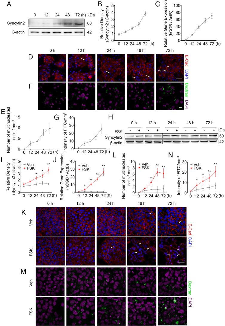
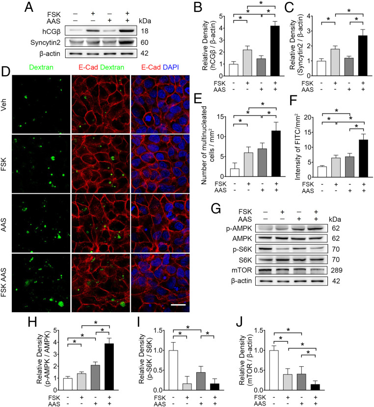
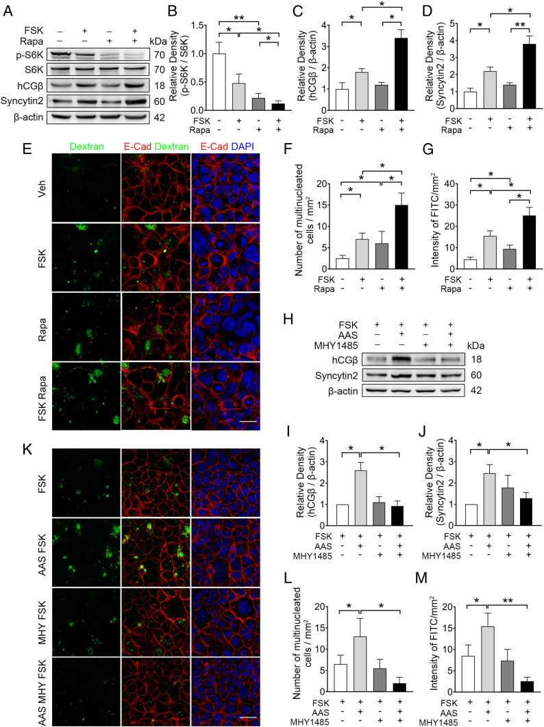
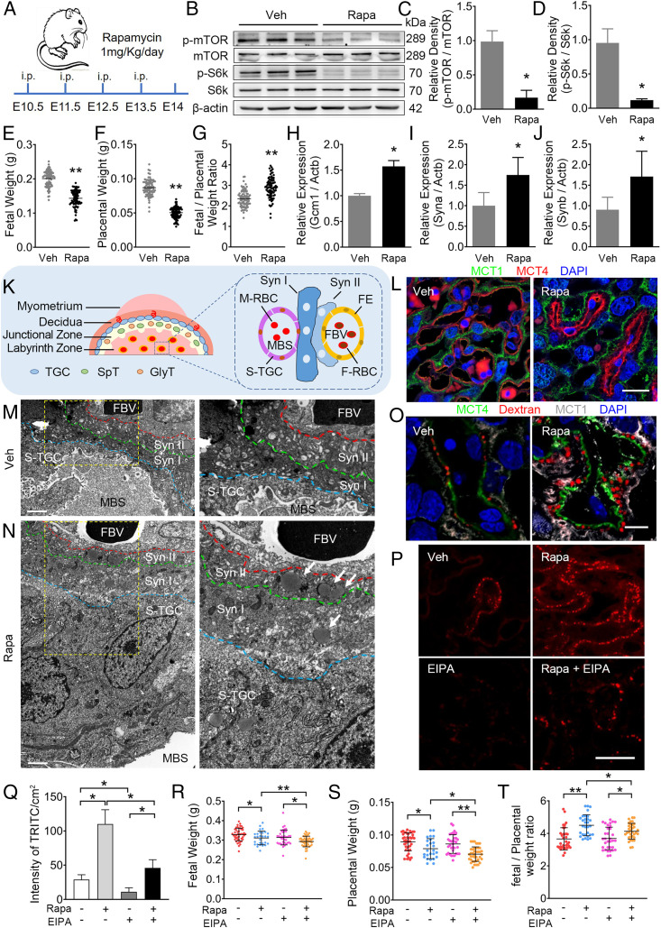
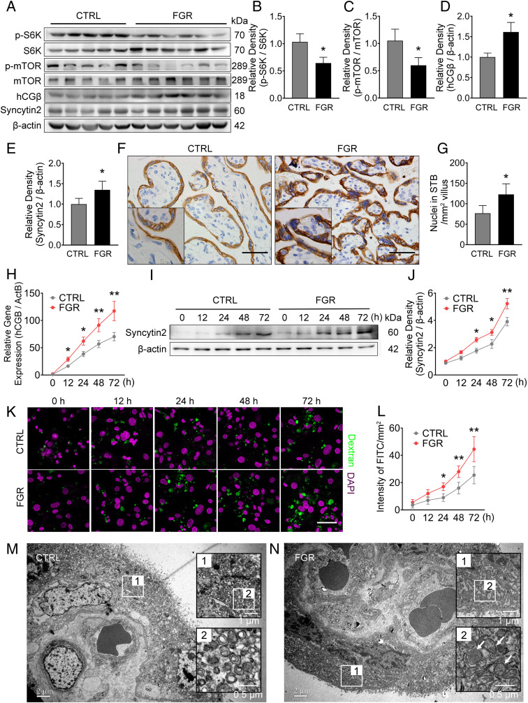
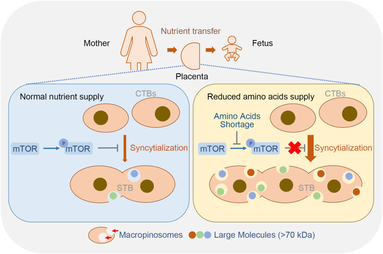
During pregnancy, the appropriate allocation of nutrients between the mother and the fetus is dominated by maternal-fetal interactions, which is primarily governed by the placenta. The syncytiotrophoblast (STB) lining at the outer surface of the placental villi is directly bathed in maternal blood and controls feto-maternal exchange. The STB is the largest multinucleated cell type in the human body, and is formed through syncytialization of the mononucleated cytotrophoblast. However, the physiological advantage of forming such an extensively multinucleated cellular structure remains poorly understood. Here, we discover that the STB uniquely adapts to nutrient stress by inducing the macropinocytosis machinery through repression of mammalian target of rapamycin (mTOR) signaling. In primary human trophoblasts and in trophoblast cell lines, differentiation toward a syncytium triggers macropinocytosis, which is greatly enhanced during amino acid shortage, induced by inhibiting mTOR signaling. Moreover, inhibiting mTOR in pregnant mice markedly stimulates macropinocytosis in the syncytium. Blocking macropinocytosis worsens the phenotypes of fetal growth restriction caused by mTOR-inhibition. Consistently, placentas derived from fetal growth restriction patients display: 1) Repressed mTOR signaling, 2) increased syncytialization, and 3) enhanced macropinocytosis. Together, our findings suggest that the unique ability of STB to undergo macropinocytosis serves as an essential adaptation to the cellular nutrient status, and support fetal survival and growth under nutrient deprivation.

Keywords: amino acid shortage; fetal growth; mTOR; macropinocytosis; placental syncytiotrophoblast.


Conflict of interest statement

The authors declare no competing interest.

Figures

Fig. 1.
Fig. 1.
Syncytialization is associated with higher levels of macropinocytosis in human trophoblasts. (AC) Western blot of Syncytin2 (A and B) and mRNA level of hCGB (C) in term PHT cells. (D and E) Immunostainings of E-cadherin (red) and DAPI (blue) in primary CTBs from normal term placenta, showing spontaneous syncytialization. (D) Representative image of syncytialized trophoblasts. White arrows, multinucleated trophoblasts. (Scale bar, 40 μm.) (E) Quantification of multinucleated cell. E-cadherin staining outlines cell boundary. The number of multinucleated cells was calculated by counting total number of cells with at least two nuclei in five randomly selected fields per sample, three replicates per group. The results are presented as mean ± SD per square millimeter per group. (F and G) Representative FITC-dextran uptake, reflecting macropinocytosis in primary trophoblasts. (F) Representative confocal images showing the intracellular accumulation of fluorescent puncta after 30-min incubation in medium that was supplemented by >70 kDa FITC-dextran. Green and magenta signals indicate FITC-dextran and DAPI, respectively. The macropinocytosis activity was analyzed using ImageJ software (NIH). The “Smooth” feature in ImageJ was employed after background subtraction and before threshold adjustments. The area of FITC-labeled macropinosomes was determined using the “Analyze Particles” feature in ImageJ, and the intensity of FITC of macropinosomes was computed by the mean area of five randomly selected fields per slide, three slides per group (47). (Scale bar, 40 μm.) (G) A time course of FITC intensity reflecting macropinocytosis in trophoblasts. (H–J) Western blot of Syncytin2 (H and I) and mRNA (RT-qPCR) of hCGB (J) in 20 μM FSK exposed BeWo cells. (K) Representative images of 20 μM FSK-induced syncytialization of BeWo cells, with the white arrows indicating multinucleated cells. (Scale bar, 40 μm.) (L) Quantification of multinucleated cell in K. (M) Representative FITC-dextran uptake images, measuring macropinocytosis in FSK exposed BeWo cells. (Scale bar, 40 μm.) (N) Quantification of FITC intensity. The data are shown as mean ± SD, and analysis was carried out by two-tailed t test based at least three independent experiments. *P < 0.05; **P < 0.01.
Fig. 2.
Fig. 2.
AAS simultaneously promotes syncytialization and macropinocytosis in BeWo cells. (A–C) Western blots (A) and corresponding quantification (B and C) of hCGβ and syncytin2 in BeWo cells cultured in AAS, with concentrations of Lys, Glu, and Arg at one-eighth of the normal levels with or without 20 μM FSK. (D) Representative immunostaining of dextran (green), E-cadherin (red), and DAPI (blue) in BeWo cells cultured in conditions detailed. (Scale bar, 40 μm.) (E and F) Quantification of multinucleated cell and FITC intensity. (G–J) Western blots (G) and corresponding quantification (H–J) of p-AMPK, AMPK, p-S6K, S6K, and mTOR in BeWo cells cultured in the AAS condition with or without 20 μM FSK. (H–J) Semiquantification of the relative density of p-AMPK/AMPK, p-S6K/S6K, and mTOR. Data were shown as mean ± SD and analyzed by one-way ANOVA test and Tukey–Kramer multiple comparison test based on at least three independent experiments. *P < 0.05.
Fig. 3.
Fig. 3.
mTOR inhibition is necessary for the induction of syncytialization and macropinocytosis by FSK in BeWo cells. (A–D) Western blots (A) and corresponding semiquantification (B–D) of p-S6K, S6K, hCGβ,and Syncytin2 in BeWo cells exposed to 100 nM Rapa, with or without 20 μM FSK. (B–D) Semiquantification of p-S6K/S6K, hCGβ, and Syncytin2. (E and F) Representative immunostaining of E-cadherin (red) and DAPI (blue) in BeWo cells cultured in the indicated conditions in E. (Scale bar, 40 μm.) (F) Quantification of multinucleated BeWo cell. (E and G) Representative FITC-dextran (green) uptake in BeWo cells, cultured as detailed in E. (G) Quantification of FITC intensity. (H–J) Western blots (H) and corresponding quantification (I and J) of hCGβ and syncytin2 in BeWo cells cultured in AAS media, exposed to 20 μM FSK, with or without 10 μM MHY1485. (K and L) Representative immunostaining of E-cadherin (red) and DAPI (blue) in BeWo cells cultured as detailed in K. (Scale bar, 40 μm.) (L) Quantification of multinucleated BeWo cell. (K and M) Representative FITC-dextran (green) uptake experiment in BeWo cells cultured in indicated conditions in K. (M) Quantification of FITC intensity. The data are shown as mean ± SD and analyzed by one-way ANOVA test and Tukey–Kramer multiple comparison test based on at least three independent experiments. *P < 0.05; **P < 0.01.
Fig. 4.
Fig. 4.
Inhibition of mTOR signaling by Rapa in pregnant mice leads to enhanced trophoblast syncytialization and macropinocytosis. (A) A schematic depiction of the experimental settings. Pregnant CD-1 mice were intraperitoneally injected with 1 mg/kg Rapa or vehicle (DMSO) daily from E10.5 to E13.5, and the mice were killed at E14. (BD) Western blots (B) and the corresponding quantification (C and D) of p-mTOR, mTOR, p-S6k, and S6k in mouse placentas treated with vehicle (n = 10) or Rapa (n = 10). (C and D) Semiquantification of p-mTOR/mTOR and p-S6k/S6k. (EG) FW (E), PW (F), and FW/PW weight ratio (G) in vehicle or Rapa dams. (HJ) mRNA levels of STB makers, Gcm1 (H), Syna (I), and Synb (J), in placentas from mice treated with vehicle or Rapa. (K) Schematic depiction of the mouse placenta. FE, fetal endothelium; FBV, fetal blood vessel; F-RBC, fetal red blood cell; GlyT, glycogen trophoblast; MBS, maternal blood sinus; M-RBC, maternal red blood cell; SpT, spongiotrophoblast; S-TGC, sinusoidal trophoblast giant cell; Syn I, the first layer of STB; Syn II, the second layer of STB; TGC, trophoblast giant cell. (L) Immunofluorescent staining of MCT1 (green), MCT4 (red) in the indicated placentas. Scale bar, 20 μm. (M and N) Representative TEM images of STBs in mouse placentas treated with vehicle (M, n = 6) or Rapa (N, n = 6). Right in M and N are the corresponding higher magnification images of the yellow frame Insets (Left). White arrows, large vesicles (0.2 to ∼5 μm in diameter, indicating macropinosomes) observed in Syn II of Rapa-treated mice. (Scale bars, 1 μm; magnification used in enlargements, 2×) (O) Three mice per group were subjected to single-injection of 2 mg/kg TRITC-dextran (intravenously) 3 min before being killed. Representative confocal images of TRITC-dextran (red) uptake in mouse STBs illustrated by MCT4 (green) or MCT1 (white) in vehicle or Rapa. (Scale bar, 20 μm.) (P–T) Pregnant CD-1 mice were injected from E10.5 to E13.5 with vehicle (DMSO, intraperitoneally and/or intravenously, n = 5), 1 mg/kg/d of Rapa (intraperitoneally, n = 5), 1 mg/kg/d of EIPA (intravenously, n = 5), and combination of Rapa and EIPA (n = 5), respectively. The placenta tissues were collected at E14. (P) Representative staining of TRITC-dextran (red) in the indicated E14 placentas of vehicle (DMSO, n = 3), Rapa- (n = 3), EIPA- (n = 3), and Rapa+EIPA- (n = 3) treated mice. (Scale bar, 40 μm.) (Q) Quantification of TRITC intensity in the indicated mouse placentas. (R–T) FW (R), PW (S), and FW/PW weight ratio (T) in vehicle-, Rapa-, EIPA-, and Rapa+EIPA-treated groups. The data are shown as mean ± SD, and the analysis was carried out by two-tailed t test (CJ) or one-way ANOVA test and Tukey–Kramer multiple-comparison test (Q–S). *P < 0.05; **P < 0.01.
Fig. 5.
Fig. 5.
Syncytialization, macropinocytosis activity, and mTOR signaling in normal or FGR human placentas. Sixteen control and 14 FGR placentas were collected and analyzed. (A–E) Western Blots (A) and corresponding quantification (B–E) of p-S6K, S6K, p-mTOR, mTOR, hCGβ, and Syncytin2 in control and FGR placentas. (F and G) Quantification of the number of nuclei in STB of control and FGR placentas based on the immunohistochemistry staining of Cytokeratin 7 (F, higher magnification of images are displayed as Insets). (G) Quantification of nuclear number in STB. Three placentas from each group were included, and three slides per sample were subjected to immunohistochemistry. The number of nuclei in the STB was counted in five random fields of each slide and presented as nuclear number per square millimeter of villous area. The results are presented as mean ± SD, and statistically analyzed by two-tailed t test. (Scale bars, 400 μm; magnification used in enlargements in F, 2×) (H–J) mRNA level of hCGB (H) and Western blot of syncytin2 (I and J) in primary cultured trophoblasts derived from control or FGR placentas. (K) Representative images of FITC-dextran uptake indicating macropinocytosis in PHT cells derived from control or FGR placenta, with green and magenta signal indicating FITC-dextran and DAPI, respectively. (Scale bar, 20 μm.) (L) Quantification of FITC intensity. (M and N) Representative TEM images of STBs from normal pregnancy (M, n = 5) vs. FGR (N, n = 5) placentas. The panel in black frames are regional magnification of the corresponding white frames, with white arrows indicating large vesicles (0.2 to ∼5 μm in diameter, indicating macropinosomes) observed in the STBs of the FGR placenta. The data are shown as mean ± SD, analyzed by two-tailed t test based on the results from at least three independent experiments. *P < 0.05; **P < 0.01.
Fig. 6.
Fig. 6.
A model of macropinocytosis-mediated adaptation to nutritional stress in STB. Macropinocytosis in trophoblasts positively correlates with syncytialization. The syncytialized placental trophoblasts can efficiently uptake large molecules (>70 kDa) by the macropinocytosis machinery, which is enhanced during reduced amino acid supply. AAS induces trophoblast syncytialization and activates macropinocytosis by inhibiting mTOR phosphorylation and activation.

References

    1. Anin S. A., Vince G., Quenby S., Trophoblast invasion. Hum. Fertil. (Camb.) 7, 169–174 (2004). - PubMed
    1. Lin G., et al. , Improving amino acid nutrition to prevent intrauterine growth restriction in mammals. Amino Acids 46, 1605–1623 (2014). - PubMed
    1. Zhang S., et al. , Placental adaptations in growth restriction. Nutrients 7, 360–389 (2015). - PMC - PubMed
    1. Mandruzzato G. et al.; WAPM , Intrauterine restriction (IUGR). J. Perinat. Med. 36, 277–281 (2008). - PubMed
    1. McIntire D. D., Bloom S. L., Casey B. M., Leveno K. J., Birth weight in relation to morbidity and mortality among newborn infants. N. Engl. J. Med. 340, 1234–1238 (1999). - PubMed

Publication types

MeSH terms

LinkOut - more resources