. 2021 Jan-Feb;38(1):41-52.
doi: 10.4103/lungindia.lungindia_326_19.

Percutaneous core needle biopsy in the diagnosis of lung lesions: An experience on 280 consecutive cases from a university hospital in southern India

Affiliations

Percutaneous core needle biopsy in the diagnosis of lung lesions: An experience on 280 consecutive cases from a university hospital in southern India

Madhavi Parigi et al. Lung India. 2021 Jan-Feb.

Abstract

Context: Percutaneous needle biopsy of lung (PCNBL) is advantageous over bronchoscopic biopsies to obtain adequate sample for peripheral lung lesions.

Objective: The objective was to evaluate the diagnostic yield of image-guided PCNBL in the diagnosis of lung lesions and to classify lung carcinomas as per the recently proposed International Association for the Study of Lung Cancer (IASLC)/American Thoracic Society/European Respiratory Society classification for small biopsies modified and adopted by the World Health Organization, 2015.

Materials and methods: A total of 280 image-guided PCNBL were analyzed. The radiological findings and routine hematoxylin and eosin (H&E)-stained sections along with immunohistochemistry (IHC) were analyzed in all the cases. Molecular testing was done depending on tissue diagnosis and availability.

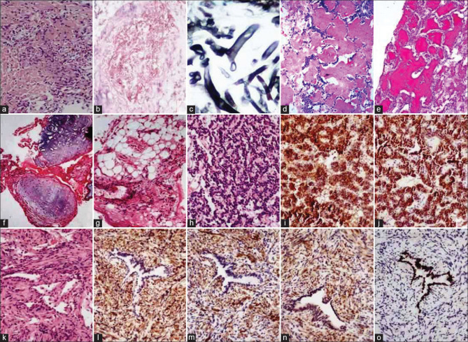
Results: Majority (81%) were diagnosed as malignant lesions, with adenocarcinoma (ADC) being the most common. More than 70% were diagnosed on H&E morphology alone, with thirty cases requiring IHC to categorize as ADC. Nearly 60% were categorized as squamous cell carcinoma on morphology alone and the rest required IHC. Though TTF1 showed higher sensitivity than napsin A, the latter is more specific. Both p63 and p40 were found to be highly sensitive for squamous cell carcinoma, but p40 was more specific than p63. Epidermal growth factor receptor could be evaluated on 94.4% of ADC samples, indicating good yield for molecular testing.

Conclusion: PCNBL yields adequate sampling for tissue diagnosis and ancillary testing with minimal complications. The use of IHC markers reduces the number of non-small-cell not otherwise specified cases significantly.

Keywords: Immunohistochemistry; molecular tests; percutaneous needle biopsy of lung.


Conflict of interest statement

None

Figures

Figure 1
Figure 1
(a) Necrotizing granulomatous inflammation (H and E; ×100). (b) Pulmonary zygomycosis showing infarcted lung tissue with fungal hyphae (H and E; ×400). (c) Broad aseptate hyphae highlighted on GMS stain (GMS; ×400). (d and e) Pulmonary alveolar proteinosis showing alveolar spaces filled with acellular eosinophilic material highlighted on PAS stain (H and E and PAS; ×400). (f and g) Pulmonary chondroid hamartoma showing lobules of hyaline cartilage and mature adipose tissue (H and E; ×100). (h) Typical carcinoid showing interconnecting cords of cells with a uniform round nucleus (H and E; ×100). (i and j) Cells showing diffuse strong positivity for synaptophysin and chromogranin (poly HRP; ×100). (k-o) Inflammatory myofibroblastic tumor showing interlacing fascicles of spindle cells with entrapped alveoli. Cells are positive for vimentin, smooth muscle actin, and anaplastic lymphoma kinase and negative for TTF1 (H and E, ×100 [k] and poly HRP ×100 [l-o])
Figure 2
Figure 2
(a-f) Squamous cell carcinoma: (a) Nests of polygonal cells with nuclear pleomorphism and hyperchromasia (H and E; ×100). (b-f) On immunohistochemistry, cells are negative for TTF1 and napsin A but positive for p63, p40, and CK5/6 (poly HRP; ×100). (g-l) Non-small-cell carcinoma NOS: (g) Sheets of undifferentiated cells with foci of necrosis (H and E; ×100). (h-l) On immunohistochemistry, the tumor cells are negative for TTF1, Napsin-A, p-40, and CK5/6 but positive for pan-CK (poly HRP; ×100). (m-r) Small-cell carcinoma: (m) Sheets of small cells with hyperchromatic nucleus and inconspicuous nucleoli (H and E; ×100). (n-r) On immunohistochemistry, the tumor cells are positive for chromogranin, synaptophysin, and TTF1 but negative for P63 (poly HRP; ×100)
Figure 3
Figure 3
(a-f) Lepidic adenocarcinoma: (a) Cuboidal cells lining the preexisting alveolar spaces (H and E; ×100). (b-f) Tumor cells are positive for TTF1, napsin A, CK7, and p63 and negative for p40 (poly HRP; ×100). (g-l) Invasive mucinous adenocarcinoma: (g) Acinar structures filled with mucin (H and E; ×100). (h-l) On immunohistochemistry, cells are positive for TTF1, napsin A, and CK7 and negative for CK20. P63 shows positive staining in the tumor cells (poly HRP; ×100). (m-r) Mucinous adenocarcinoma with lepidic pattern: (m and n) Columnar cells with basally located nuclei and intracytoplasmic mucin lining the preexisting alveolar spaces (H and E; ×40 and ×100). (o-r) On immunohistochemistry, cells are negative for TTF1 and napsin A and positive for CK20 (focal) and CK7 (poly HRP; ×100). (s) Micropapillary adenocarcinoma showing micropapillae without central fibrovascular core (H and E; ×100). (t-x) Papillary adenocarcinoma: (t) Papillae lined with cuboidal-to-columnar cells lining the papillae (H and E; ×100). (u-x) Cells show positivity for TTF1 and napsin A and p63 but negative for p40 (poly HRP; ×100)
Figure 4
Figure 4
(a-e) Metastatic endometrioid carcinoma: (a) Infiltrating tubular glands lined by tall columnar cells (H and E; ×100). (b-e) On immunohistochemistry, the tumor cells are negative for TTF1 and napsin A and positive for estrogen receptor and vimentin (poly HRP; ×100). (f-j) Anaplastic large-cell lymphoma: (f) Diffuse sheets of small and large lymphoid cells admixed with plasma cells and eosinophils (H and E; ×100). (g-j) Cells are negative for pan CK and TTF1 and positive for LCA and CD30 (poly HRP; ×100 [g-i] and ×400 [j]). (k-o) Synovial sarcoma: (k) Sheets and fascicles of spindle cells (H and E; ×100). (l-o) Cells are positive for Bcl2, pan-CK, and EMA and negative for CD34 (poly HRP; ×100)

References

    1. Cardella JF, Bakal CW, Bertino RE, Burke DR, Drooz A, Haskal Z, et al. Quality improvement guidelines for image-guided percutaneous biopsy in adults. J Vasc Interv Radiol. 2003;14:S227–30. - PubMed
    1. Nikolaidis P, van Sonnenberg E, Haddad ZK, Chen YH, Zou KH, Tuncali K, et al. Practice patterns of nonvascular interventional radiology procedures at academic centers in the United States? Acad Radiol. 2005;12:1475–82. - PubMed
    1. Richardson CM, Pointon KS, Manhire AR, Macfarlane JT. Percutaneous lung biopsies: A survey of UK practice based on 5444 biopsies. Br J Radiol. 2002;75:731–5. - PubMed
    1. Stanley JH, Fish GD, Andriole JG, Gobien RP, Betsill WL, Laden SA, et al. Lung lesions: Cytologic diagnosis by fine-needle biopsy. Radiology. 1987;162:389–91. - PubMed
    1. Lucidarme O, Howarth N, Finet JF, Grenier PA. Intrapulmonary lesions: Percutaneous automated biopsy with a detachable, 18-gauge, coaxial cutting needle. Radiology. 1998;207:759–65. - PubMed