

Roles of Exosomes in Ocular Diseases
- PMID: 33402823
- PMCID: PMC7778680
- DOI: 10.2147/IJN.S277190
Roles of Exosomes in Ocular Diseases
Abstract
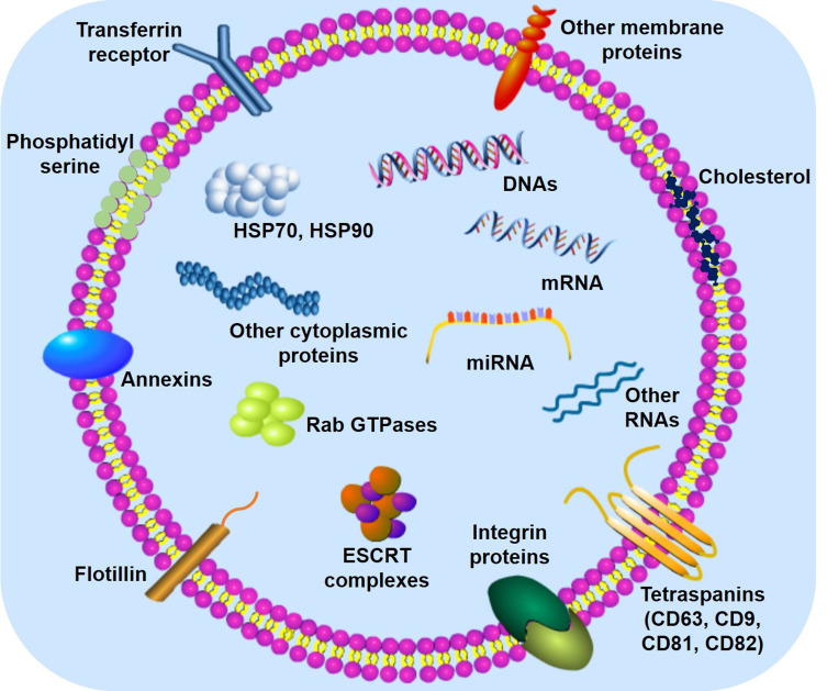
Exosomes, nanoscale vesicles with a diameter of 30 to 150 nm, are composed of a lipid bilayer, protein, and genetic material. Exosomes are secreted by virtually all types of cells in the human body. They have key functions in cell-to-cell communication, immune regulation, inflammatory response, and neovascularization. Mounting evidence indicates that exosomes play an important role in various diseases, such as cancer, cardiovascular diseases, and brain diseases; however, the role that exosomes play in eye diseases has not yet been rigorously studied. This review covers current exosome research as it relates to ocular diseases including diabetic retinopathy, age-related macular degeneration, autoimmune uveitis, glaucoma, traumatic optic neuropathies, corneal diseases, retinopathy of prematurity, and uveal melanoma. In addition, we discuss recent advances in the biological functions of exosomes, focusing on the toxicity of exosomes and the use of exosomes as biomarkers and drug delivery vesicles. Finally, we summarize the primary considerations and challenges to be taken into account for the effective applications of exosomes.
Keywords: drug delivery; exosomes; extracellular vesicles; ocular diseases; retina.
© 2020 Liu et al.
Conflict of interest statement
The authors report no conflicts of interest for this work.
Figures


Similar articles
-
Recent advances of exosomes in immune-mediated eye diseases.Stem Cell Res Ther. 2019 Aug 30;10(1):278. doi: 10.1186/s13287-019-1372-0. Stem Cell Res Ther. 2019. PMID: 31470892 Free PMC article. Review.
-
Intraocular Exosomes in Eye Diseases.Curr Mol Med. 2022;22(6):540-548. doi: 10.2174/1566524021666210901122948. Curr Mol Med. 2022. PMID: 34488586 Review.
-
Extracellular vesicles in degenerative retinal diseases: A new therapeutic paradigm.J Control Release. 2024 Jan;365:448-468. doi: 10.1016/j.jconrel.2023.11.035. Epub 2023 Dec 2. J Control Release. 2024. PMID: 38013069 Review.
-
Circulating exosomes in ophthalmic disease: novel carriers of biological information circulating exosomes in ophthalmic disease.Eur Rev Med Pharmacol Sci. 2021 Mar;25(5):2172-2181. doi: 10.26355/eurrev_202103_25208. Eur Rev Med Pharmacol Sci. 2021. PMID: 33755954 Review.
-
Exosomes, extracellular vesicles and the eye.Exp Eye Res. 2022 Jan;214:108892. doi: 10.1016/j.exer.2021.108892. Epub 2021 Dec 10. Exp Eye Res. 2022. PMID: 34896308 Review.
Cited by
-
Exosomal microRNAs as potential biomarkers and therapeutic targets in corneal diseases.Mol Vis. 2024 Mar 15;30:92-106. eCollection 2024. Mol Vis. 2024. Retraction in: Mol Vis. 2024 Aug 29;30:278. PMID: 38601014 Free PMC article. Retracted. Review.
-
Target specification and therapeutic potential of extracellular vesicles for regulating corneal angiogenesis, lymphangiogenesis, and nerve repair.Ocul Surf. 2024 Oct;34:459-476. doi: 10.1016/j.jtos.2024.10.005. Epub 2024 Oct 18. Ocul Surf. 2024. PMID: 39426677 Review.
-
Nanomaterials targeting cancer stem cells to overcome drug resistance and tumor recurrence.Front Oncol. 2025 Jun 6;15:1499283. doi: 10.3389/fonc.2025.1499283. eCollection 2025. Front Oncol. 2025. PMID: 40548119 Free PMC article. Review.
-
EndMT Regulation by Small RNAs in Diabetes-Associated Fibrotic Conditions: Potential Link With Oxidative Stress.Front Cell Dev Biol. 2021 May 19;9:683594. doi: 10.3389/fcell.2021.683594. eCollection 2021. Front Cell Dev Biol. 2021. PMID: 34095153 Free PMC article. Review.
-
Lysyl oxidase like-1 deficiency in optic nerve head astrocytes elicits reactive astrocytosis and alters functional effects of astrocyte derived exosomes.Exp Eye Res. 2024 Mar;240:109813. doi: 10.1016/j.exer.2024.109813. Epub 2024 Feb 6. Exp Eye Res. 2024. PMID: 38331016 Free PMC article.
References
-
- Johnstone RM, Adam M, Hammond JR, Orr L, Turbide C. Vesicle formation during reticulocyte maturation. Association of plasma membrane activities with released vesicles (exosomes). J Biol Chem. 1987;262(19):9412–9420. - PubMed
