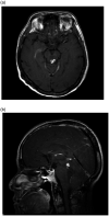
One of a kind-chordoid glioma in the fourth ventricle: a case report and literature review
- PMID: 33403125
- PMCID: PMC7739103
- DOI: 10.1177/2058460120980143
One of a kind-chordoid glioma in the fourth ventricle: a case report and literature review
Abstract
Chordoid glioma (CG) is a rare brain tumor that is known for its characteristic location in the third ventricle. A wide spectrum of radiological presentations has been described, with few common features among them. Its radiological diagnosis is mainly suggested by location. However, several cases of CG with atypical locations have been described, illustrating that CG is not limited to the third ventricle, and should be considered in the list of radiological differential diagnosis for intraventricular masses. We present here a case of CG that was found in the fourth ventricle.
Keywords: Chordoid glioma; fourth ventricle; intraventricular tumor; magnetic resonance imaging.
© The Foundation Acta Radiologica 2020.
Figures


Similar articles
-
Chordoid glioma of the third ventricle: a patient presenting with SIADH and a review of this rare tumor.Pituitary. 2016 Aug;19(4):356-61. doi: 10.1007/s11102-016-0711-8. Pituitary. 2016. PMID: 26879322 Review.
-
Chordoid Glioma with Intraventricular Dissemination: A Case Report with Perfusion MR Imaging Features.Korean J Radiol. 2016 Jan-Feb;17(1):142-6. doi: 10.3348/kjr.2016.17.1.142. Epub 2016 Jan 6. Korean J Radiol. 2016. PMID: 26798226 Free PMC article.
-
Chordoid glioma: a rare old foe but a new pathological and radiological presentation.Clin Imaging. 2021 Oct;78:160-164. doi: 10.1016/j.clinimag.2021.03.008. Epub 2021 Mar 24. Clin Imaging. 2021. PMID: 33836423
-
Chordoid Glioma of the Third Ventricle: A Case Report and a Treatment Strategy to This Rare Tumor.Front Oncol. 2020 Apr 9;10:502. doi: 10.3389/fonc.2020.00502. eCollection 2020. Front Oncol. 2020. PMID: 32328466 Free PMC article.
-
Chordoid meningioma of the third ventricle: a case report and review of the literature.Clin Neuropathol. 2011 Mar-Apr;30(2):70-4. doi: 10.5414/npp30070. Clin Neuropathol. 2011. PMID: 21329615 Review.
Cited by
-
Magnetic resonance imaging of a third ventricular chordoid glioma.Radiol Case Rep. 2021 Jun 8;16(8):1941-1945. doi: 10.1016/j.radcr.2021.04.074. eCollection 2021 Aug. Radiol Case Rep. 2021. PMID: 34149979 Free PMC article.
References
-
- Brat DJ, Scheithauer BW, Staugaitis SM, et al. Third ventricular chordoid glioma: a distinct clinicopathologic entity. J Neuropathol Exp Neurol 1998; 57:283–290. - PubMed
-
- Chen F, Cao JG, Liu XL, et al. Case Report Lateral ventricular chordoid glioma in a pediatric patient: a case report and literature review. Int J Clin Exp Med 2017; 10:8410–8414. - PubMed
-
- Jain D, Sharma MC, Sarkar C, et al. Chordoid glioma: report of two rare examples with unusual features. Acta Neurochir 2008; 150:295–300. - PubMed
Publication types
LinkOut - more resources
Full Text Sources

