

Role of CT Imaging With Three-Dimensional Maximum Intensity Projection Reconstruction in the Evaluation of Portal Vein Variants at a Tertiary Care Hospital
- PMID: 33403165
- PMCID: PMC7773306
- DOI: 10.7759/cureus.11733
Role of CT Imaging With Three-Dimensional Maximum Intensity Projection Reconstruction in the Evaluation of Portal Vein Variants at a Tertiary Care Hospital
Abstract
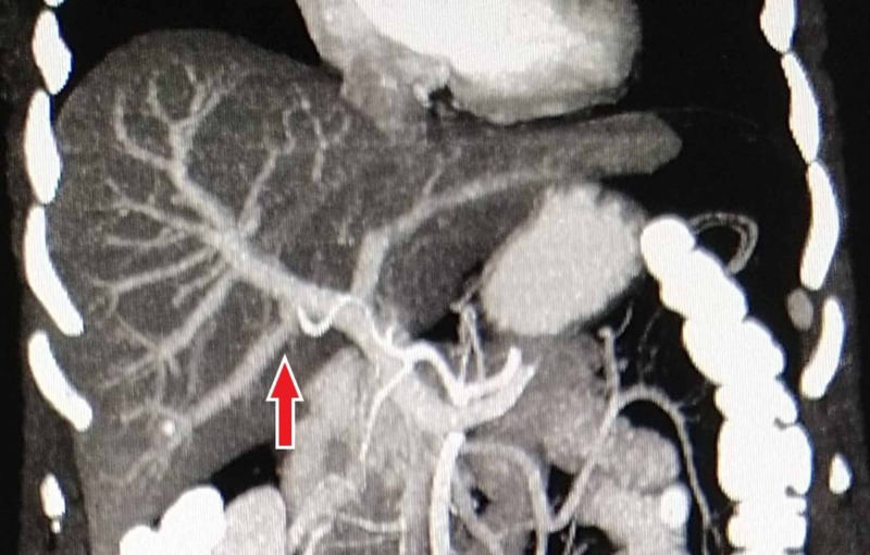
Introduction: Portal vein (PV) is the principal blood vessel transporting blood from the alimentary tract and spleen to the liver. The aim of this study is to determine the prevalence of PV anatomical variations in our population using multidetector CT with maximum intensity projection (MIP) technique at a tertiary care hospital.
Methods: This cross-sectional study was prospectively conducted from November 2018 to June 2019 in the Department of Radiology at a tertiary care hospital in Karachi. After informed consent, all the patients with no known hepatic pathology undergoing routine abdomen CT were included in this study. Patients with previous hepatic resection surgeries, undiagnosed large hepatic tumors/metastasis, and those with PV thrombosis were excluded.
Results: A total of 500 patients (256 males and 244 females) were included in the study; the mean age of female patients was relatively higher as compared to the male patients (53.80 ± 18.44 vs. 44.15 ± 19.94 years; p = 0.000). Standard PV anatomy (type 1) was found in 438 patients (87.6%). Trifurcation (type 2) occurred in 18 patients (3.6%). Right posterior portal vein as the first branch of main PV (type 3) was found in 22 patients (4.4%). A separate branch of the right portal vein (RPV) to segment VII (type 4) and separate branch of the RPV to segment VI (type 5) were found in 6 (1.2%) and 16 (3.2%) patients, respectively.
Conclusion: Our study displayed a relatively higher frequency of standard PV anatomy (type 1) compared to previous studies. We highlight the role of MIP in the analysis of hepatic venous anatomy with its utility demonstrating improved detection of variations.
Keywords: computed tomography; maximum intensity projection; portal vein variant.
Copyright © 2020, Asad Ullah et al.
Conflict of interest statement
The authors have declared that no competing interests exist.
Figures


Similar articles
-
CT virtual endoscopy for analyzing variations in the hepatic portal vein.Surg Radiol Anat. 2015 Jul;37(5):457-62. doi: 10.1007/s00276-015-1463-2. Epub 2015 Mar 25. Surg Radiol Anat. 2015. PMID: 25804700
-
Multislice CT angiography in the evaluation of hepatic vascular anatomy in potential right lobe donors.Diagn Interv Radiol. 2005 Mar;11(1):51-9. Diagn Interv Radiol. 2005. PMID: 15795845
-
Variations of the right branch of hepatic portal vein in children based on three-dimensional simulation technology.Surg Radiol Anat. 2020 Dec;42(12):1467-1473. doi: 10.1007/s00276-020-02499-3. Epub 2020 May 18. Surg Radiol Anat. 2020. PMID: 32424682
-
Portal Vein Variations, Clinical Correlation, and Embryological Explanation: A Review Article.Cureus. 2023 Mar 20;15(3):e36400. doi: 10.7759/cureus.36400. eCollection 2023 Mar. Cureus. 2023. PMID: 37090306 Free PMC article. Review.
-
Surgical Implications of Portal Vein Variations and Liver Segmentations: A Recent Update.J Clin Diagn Res. 2017 Feb;11(2):AE01-AE05. doi: 10.7860/JCDR/2017/25028.9453. Epub 2017 Feb 1. J Clin Diagn Res. 2017. PMID: 28384848 Free PMC article. Review.
Cited by
-
Examining the Branching Patterns of the Hepatis Portae Vena with Computed Tomography Images.J Clin Med. 2025 Jul 8;14(14):4835. doi: 10.3390/jcm14144835. J Clin Med. 2025. PMID: 40725529 Free PMC article.
-
Duplicated inferior vena cava-trifurcated portal vein: a rare anatomical variation encountered during Whipple procedure.J Surg Case Rep. 2023 Jan 26;2023(1):rjad014. doi: 10.1093/jscr/rjad014. eCollection 2023 Jan. J Surg Case Rep. 2023. PMID: 36727120 Free PMC article.
-
A systematic review and meta-analysis: prevalence and clinical implications of anatomical variants of the hepatic portal vein.Sci Rep. 2024 Dec 3;14(1):30002. doi: 10.1038/s41598-024-81543-3. Sci Rep. 2024. PMID: 39622958 Free PMC article.
References
-
- Portal vein abnormalities: an imaging review. Madhusudhan KS, Vyas S, Sharma S, Srivastava DN, Gupta AK. Clin Imaging. 2018;52:70–78. - PubMed
-
- Hepatic and portal veins anatomical variants: prevalence and clinical implications on routine abdominal MDCT. Mehmood K, Sami F, Jamal M, Pervaiz A, Bahadur HS, Aziz S. http://www.pakjr.com/ojs/index.php/PJR/article/view/960 Pak J Radiol. 2018;28:224–229.
-
- Prevalence and types of main and right portal vein branching variations on MDCT. Atasoy Ç, Özyürek E. AJR Am J Roentgenol. 2006;187:676–681. - PubMed
LinkOut - more resources
Full Text Sources