

Radiation-Induced Malignant Peripheral Nerve Sheath Tumor of the Vagus Nerve Following Radiation Treatment of Cervical Paraganglioma
- PMID: 33403195
- PMCID: PMC7775188
- DOI: 10.1055/s-0040-1718408
Radiation-Induced Malignant Peripheral Nerve Sheath Tumor of the Vagus Nerve Following Radiation Treatment of Cervical Paraganglioma
Abstract
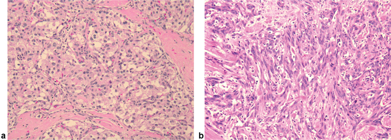
Radiation-induced sarcoma is a known but rare complication of radiation treatment for skull base paraganglioma. We present the cases of a female patient with multiple paraganglioma syndrome treated with external beam radiation treatment who presented 4 years later with a malignant peripheral nerve sheath tumor of the vagus nerve.
Keywords: glomus vagale; malignant peripheral nerve sheath tumor; paraganglioma; radiation; radiation-induced sarcoma; vagus nerve.
The Author(s). This is an open access article published by Thieme under the terms of the Creative Commons Attribution-NonDerivative-NonCommercial-License, permitting copying and reproduction so long as the original work is given appropriate credit. Contents may not be used for commercial purposes, or adapted, remixed, transformed or built upon. ( https://creativecommons.org/licenses/by-nc-nd/4.0/ ).
Conflict of interest statement
Conflict of Interest All authors certify that they have no affiliations with or involvement in any organization or entity with any financial interest (such as honoraria; educational grants; participation in speakers' bureaus; membership, employment, consultancies, stock ownership, or other equity interest; and expert testimony or patent-licensing arrangements) or nonfinancial interest (such as personal or professional relationships, affiliations, knowledge, or beliefs) in the subject matter or materials discussed in this case report.
Figures


References
-
- Galland-Girodet S, Maire J P, De-Mones E. The role of radiation therapy in the management of head and neck paragangliomas: impact of quality of life versus treatment response. Radiother Oncol. 2014;111(03):463–467. - PubMed
-
- Gilbo P, Morris C G, Amdur R J. Radiotherapy for benign head and neck paragangliomas: a 45-year experience. Cancer. 2014;120(23):3738–3743. - PubMed
-
- Liscak R, Urgosik D, Chytka T.Leksell Gamma Knife radiosurgery of the jugulotympanic glomus tumor: long-term results J Neurosurg 2014121(Suppl):198–202. - PubMed
-
- Cole J M, Beiler D. Long-term results of treatment for glomus jugulare and glomus vagale tumors with radiotherapy. Laryngoscope. 1994;104(12):1461–1465. - PubMed
-
- Hansen H S, Thomsen K A. Radiotherapy in glomus tumours (paragangliomas). A 25 year-review. Acta Otolaryngol Suppl. 1988;449:151–154. - PubMed