. 2020 Dec 16;5(51):33133-33139.
doi: 10.1021/acsomega.0c04771. eCollection 2020 Dec 29.

Decompression Process of Glycerol Shock Treatment Can Overcome Endo-Lysosomal Barriers for Intracellular Delivery

Affiliations

Decompression Process of Glycerol Shock Treatment Can Overcome Endo-Lysosomal Barriers for Intracellular Delivery

Shupeng Wang et al. ACS Omega. .

Abstract

The glycerol shock treatment has been used to improve the calcium phosphate transfection efficacy for several decades because of its high effectiveness and low toxicity. However, the mechanism of glycerol shock treatment is still obscure. In this study, the endo-lysosomal leakage assay demonstrated that the decompression process of glycerol shock treatment could enhance endo-lysosomal membrane permeabilization, which resulted in facilitating endo-lysosomal escape for effective intracellular delivery. The enhanced decompression treatment derived from glycerol shock treatment could increase the change of osmotic pressure further, which showed higher efficacy for intracellular delivery. Herein, we speculated that the endo-lysosomal swelling originated from the decompression process of glycerol shock treatment could cause endo-lysosomal damage.


Conflict of interest statement

The authors declare no competing financial interest.

Figures

Scheme 1
Scheme 1. Schematic Illustration Showing the Glycerol Shock Treatment for Intracellular Delivery
Figure 1
Figure 1
Transfection efficacy of CaP transfection in Hela and 5637 cells. Comparison of glycerol shock and NaCl shock treatment on transfection efficacy in (A,B) Hela cells and (C) 5637 cells after 48 h, respectively. Control is the group treated with 1× PBS. Scale bar, 200 μm.
Figure 2
Figure 2
Effect on hypertonic shock treatment on endo-lysosomes. (A) CLSM images of Hela cells treated with CaP/DNA for 4 h before a 15% glycerol or 6% NaCl shock, respectively. Control is the group treated with 1× PBS. Scale bar: 20 μm. (B) Schematic representation of the endo-lysosomal leakage assay. The permeabilization of endo-lysosomes allows the entry of cytosolic mAG-Gal3 proteins, leading to bright fluorescent spots. (C) Hela-mAG-Gal3 cells treated with a 15% glycerol shock. Glycerol shock treatment made mAG-Gal3 accumulate in ruptured endo-lysosomes, and bright fluorescent spots showed high colocalization with endo-lysosomes. Scale bar: 10 μm. (D) Intensity of red and green fluorescence on the arrow line in (C), mAG-Gal3: green, LysoTracker: red. Scale bar: 20 μm.
Figure 3
Figure 3
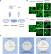
Endo-lysosomal leakage assay. (A) Schematic representation of a hypertonic shock. A hypertonic shock includes two steps. The first step is from isotonicity to hypertonicity. The second step is to return to isotonicity. (B) Fluorescence images of Hela-mAG-Gal3 cells treated with a 15% glycerol shock or a 6% NaCl shock. The decompression process enhanced endo-lysosomal membrane permeabilization. The arrows indicate ruptured endo-lysosomes. Scale bar: 50 μm. (C) CLSM images of Hela-mAG-Gal3 cells treated with hypertonic shock treatment. Scale bar: 20 μm. (D) Proposed mechanisms of decompression treatment. The dark background represents the hypertonic state; the light background represents the hypotonic state. The hypertonic solution can cause cells to lose water to shrink. Endo-lysosomes are also affected by the cell’s hyperosmotic environment, causing dehydration and shrinking. However, endo-lysosome still appeared to maintain membrane integrity. Once cells return to an isotonic solution, endo-lysosomes will swell because of the influx of water; eventually, endo-lysosomes rupture.
Figure 4
Figure 4
Enhanced decompression treatment. (A) Schematic representation of enhanced decompression treatment. Cells and endo-lysosomes will lose water to shrink in a hypertonic environment. The exchange from a hypertonic solution to an isotonic solution will lead to an increase in the influx of water. The influx of water makes the endo-lysosome swell to rupture. The exchange from a hypertonic solution to a hypotonic solution makes more water rush into endo-lysosomes, which could be damaged by more intense swelling. (B) Transfection efficacy of CaP transfection with enhanced decompression treatment in Hela cells. (C) Effects of enhanced decompression treatment on cell viability.
Figure 5
Figure 5
Decompression treatment in chitosan/DNA delivery. (A) Fluorescence images of chitosan/DNA delivery combined with decompression treatment in Hela cells. Scale bar: 200 μm. (B) Transfection efficacy of chitosan/DNA delivery combined with decompression treatment in Hela cells.

Similar articles

References

    1. Graham F. L.; van der Eb A. J. A new technique for the assay of infectivity of human adenovirus 5 DNA. Virology 1973, 52, 456–467. 10.1016/0042-6822(73)90341-3. - DOI - PubMed
    1. Jordan M.; Schallhorn A.; Wurm F. M. Transfecting mammalian cells: optimization of critical parameters affecting calcium-phosphate precipitate formation. Nucleic Acids Res. 1996, 24, 596–601. 10.1093/nar/24.4.596. - DOI - PMC - PubMed
    1. Jordan M.; Wurm F. Transfection of adherent and suspended cells by calcium phosphate. Methods 2004, 33, 136–143. 10.1016/j.ymeth.2003.11.011. - DOI - PubMed
    1. Batard P.; Jordan M.; Wurm F. Transfer of high copy number plasmid into mammalian cells by calcium phosphate transfection. Gene 2001, 270, 61–68. 10.1016/s0378-1119(01)00467-x. - DOI - PubMed
    1. Kumar P.; Nagarajan A.; Uchil P. D. Calcium Phosphate-Mediated Transfection of Eukaryotic Cells with Plasmid DNAs. Cold Spring Harbor Protoc. 2019, 2019, pdb.prot095430.10.1101/pdb.prot095430. - DOI - PubMed