

RNA-Seq identifies genes whose proteins are upregulated during syncytia development in murine C2C12 myoblasts and human BeWo trophoblasts
- PMID: 33403800
- PMCID: PMC7786548
- DOI: 10.14814/phy2.14671
RNA-Seq identifies genes whose proteins are upregulated during syncytia development in murine C2C12 myoblasts and human BeWo trophoblasts
Abstract
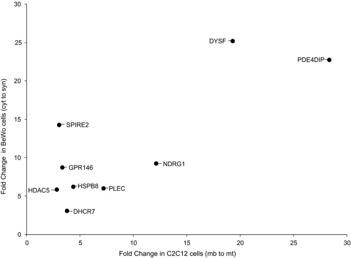
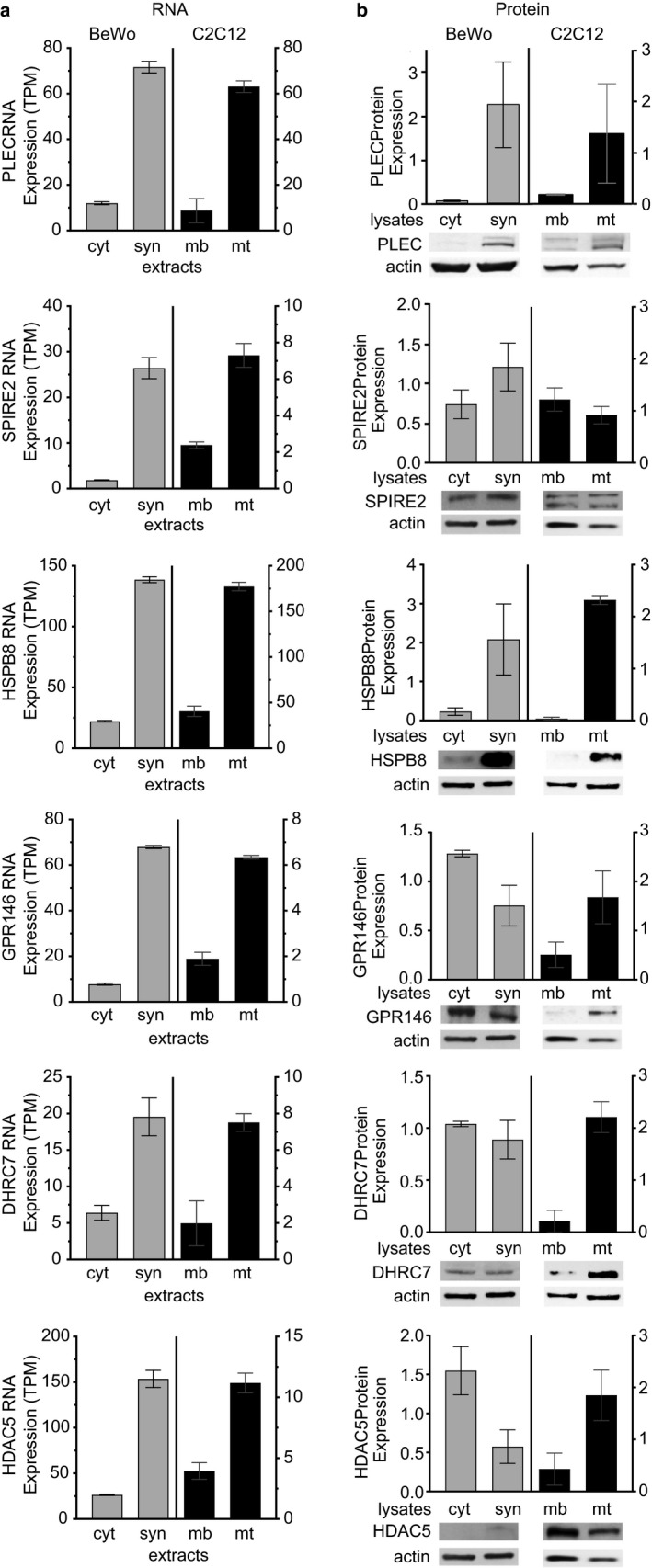
The fusion of villous cytotrophoblasts into the multinucleated syncytiotrophoblast is critical for the essential functions of the mammalian placenta. Using RNA-Seq gene expression, quantitative protein expression, and siRNA knockdown we identified genes and their cognate proteins which are similarly upregulated in two cellular models of mammalian syncytia development (human BeWo cytotrophoblast to syncytiotrophoblast and murine C2C12 myoblast to myotube). These include DYSF, PDE4DIP, SPIRE2, NDRG1, PLEC, GPR146, HSPB8, DHCR7, and HDAC5. These findings provide avenues for further understanding of the mechanisms underlying mammalian placental syncytiotrophoblast development.
Keywords: cell fusion; placenta; syncytialization.
© 2021 The Authors. Physiological Reports published by Wiley Periodicals LLC on behalf of The Physiological Society and the American Physiological Society.
Conflict of interest statement
None of the authors has any financial conflict or conflict of interest.
Figures


Similar articles
-
RNA-Seq identifies genes whose proteins are transformative in the differentiation of cytotrophoblast to syncytiotrophoblast, in human primary villous and BeWo trophoblasts.Sci Rep. 2018 Mar 23;8(1):5142. doi: 10.1038/s41598-018-23379-2. Sci Rep. 2018. PMID: 29572450 Free PMC article.
-
Involvement of nephrin in human placental trophoblast syncytialization.Reproduction. 2015 Apr;149(4):339-46. doi: 10.1530/REP-14-0424. Epub 2015 Jan 22. Reproduction. 2015. PMID: 25614620
-
Isolation, purification and in vitro differentiation of cytotrophoblast cells from human term placenta.Reprod Biol Endocrinol. 2015 Jul 9;13:71. doi: 10.1186/s12958-015-0070-8. Reprod Biol Endocrinol. 2015. PMID: 26156160 Free PMC article.
-
Scrutinising the regulators of syncytialization and their expression in pregnancy-related conditions.Mol Cell Endocrinol. 2016 Jan 15;420:180-93. doi: 10.1016/j.mce.2015.11.010. Epub 2015 Nov 14. Mol Cell Endocrinol. 2016. PMID: 26586208 Review.
-
How trophoblasts fuse: an in-depth look into placental syncytiotrophoblast formation.Cell Mol Life Sci. 2022 Jul 20;79(8):433. doi: 10.1007/s00018-022-04475-z. Cell Mol Life Sci. 2022. PMID: 35859055 Free PMC article. Review.
Cited by
-
Charting the Dynamic Trophoblast Plasma Membrane Identifies LYN As a Functional Regulator of Syncytialization.ACS Chem Biol. 2024 Oct 18;19(10):2220-2231. doi: 10.1021/acschembio.4c00443. Epub 2024 Sep 17. ACS Chem Biol. 2024. PMID: 39289808 Free PMC article.
-
Regulation of TFEB in human placental Cytotrophoblasts and Syncytiotrophoblasts.Physiol Rep. 2025 May;13(10):e70383. doi: 10.14814/phy2.70383. Physiol Rep. 2025. PMID: 40415650 Free PMC article.
-
Cost-Effective Bioimpedance Spectroscopy System for Monitoring Syncytialization In Vitro: Experimental and Numerical Validation of BeWo Cell Fusion.Micromachines (Basel). 2024 Dec 18;15(12):1506. doi: 10.3390/mi15121506. Micromachines (Basel). 2024. PMID: 39770259 Free PMC article.
-
Placental Gene Transcript Proportions are Altered in the Presence of In Utero Arsenic and Cadmium Exposures, Genetic Variants, and Birth Weight Differences.Front Genet. 2022 May 13;13:865449. doi: 10.3389/fgene.2022.865449. eCollection 2022. Front Genet. 2022. PMID: 35646058 Free PMC article.
-
Maternal and Fetal Genetic Variation in Vitamin D Metabolism and Umbilical Cord Blood 25-Hydroxyvitamin D.J Clin Endocrinol Metab. 2022 Jul 14;107(8):e3403-e3410. doi: 10.1210/clinem/dgac263. J Clin Endocrinol Metab. 2022. PMID: 35474389 Free PMC article.
References
-
- Azar, C. , Valentine, M. , Trausch‐Azar, J. , Druley, T. , Nelson, D. M. , & Schwartz, A. L. (2018). RNA‐Seq identifies genes whose proteins are transformative in the differentiation of cytotrophoblast to syncytiotrophoblast, in human primary villous and BeWo trophoblasts. Scientific Reports. Springer, US, 8, 5142 10.1038/s41598-018-23379-2 - DOI - PMC - PubMed
-
- Blaise, S. , de Parseval, N. , Benit, L. , & Heidmann, T. (2003). Genomewide screening for fusogenic human endogenous retrovirus envelopes identifies syncytin 2, a gene conserved on primate evolution. Proceedings of the National Academy of Sciences, 100(22), 13013–13018. 10.1073/pnas.2132646100 - DOI - PMC - PubMed
-
- Blond, J.‐L. , Lavillette, D. , Cheynet, Valérie , Bouton, O. , Oriol, G. , Chapel‐Fernandes, S. , …, Cosset, F.‐L. (2000). An envelope glycoprotein of the human endogenous retrovirus HERV‐W is expressed in the human placenta and fuses cells expressing the type D mammalian retrovirus receptor. Journal of Virology, 74(7), 3321–3329. 10.1128/JVI.74.7.3321-3329.2000 - DOI - PMC - PubMed
Publication types
MeSH terms
Grants and funding
LinkOut - more resources
Full Text Sources
Other Literature Sources
Miscellaneous
