Review
. 2021 Jan;23(1):33-49.
doi: 10.1177/1098612X20979509.

Feline abdominal ultrasonography: what's normal? what's abnormal? The adrenal glands

Affiliations
Review

Feline abdominal ultrasonography: what's normal? what's abnormal? The adrenal glands

Sally Griffin. J Feline Med Surg. 2021 Jan.

Abstract

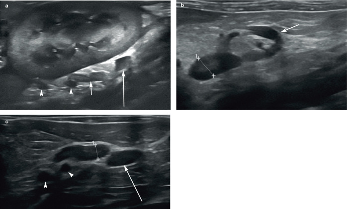
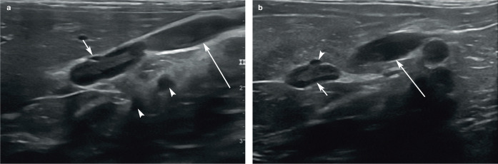
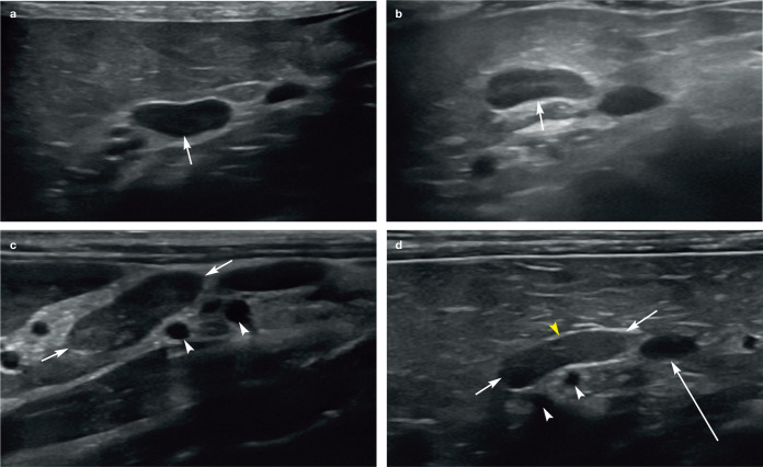
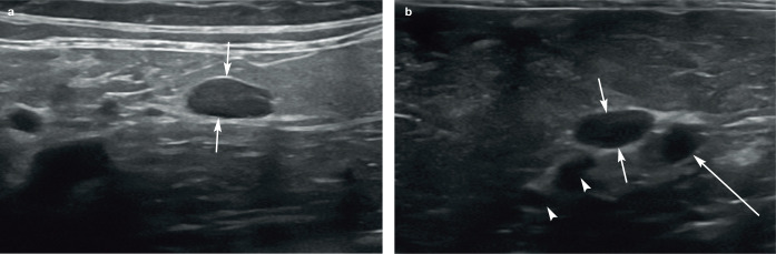
Practical relevance: Abdominal ultrasound plays a vital role in the diagnostic work-up of many cats presenting to general and specialist practitioners. Ultrasound examination of the adrenal glands can provide important information pertaining to several conditions including hyperaldosteronism and hyperadrenocorticism.

Clinical challenges: Despite ultrasonography being a commonly used modality, many practitioners are not comfortable performing an ultrasound examination or interpreting the resulting images. Even for the experienced ultrasonographer, differentiating between incidental findings, such as adrenal mineralisation, and clinically significant pathological changes can be challenging.

Aim: This review, part of an occasional series on feline abdominal ultrasonography, discusses the ultrasonographic examination of the normal and diseased adrenal glands. Aimed at general practitioners who wish to improve their knowledge of and confidence in feline abdominal ultrasound, this review is accompanied by high-resolution images and videos available online as supplementary material.

Equipment: Ultrasound facilities are readily available to most practitioners, although the use of ultrasonography as a diagnostic tool is highly dependent on operator experience.

Evidence base: Information provided in this article is drawn from the published literature and the author's own clinical experience.

Keywords: Ultrasound; acromegaly; adrenal mineralisation; hyperadrenocorticism; hyperaldosteronism; hyperthyroidism; hypoadrenocorticism; phaeochromocytoma.


Conflict of interest statement

The author declared no potential conflicts of interest with respect to the research, authorship and/or publication of this article.

Figures

Figure 1
Figure 1
Images illustrating the various landmarks in relation to the left adrenal gland in three cats. (a) The adrenal gland (short arrow) is visible medial to the kidney, caudal to the coeliac and cranial mesenteric arteries (arrowheads), and cranial to the left renal artery (long arrow), which is only partly visible. The coeliac artery emerges from the aorta cranial to the cranial mesenteric artery. (b) The left renal artery (arrow) forms a hook caudal to the left adrenal gland (between the measuring calipers). (c) The coeliac and cranial mesenteric arteries (arrowheads) are shown in cross section cranial to the adrenal gland (between the measuring calipers). A portion of the left renal artery (long arrow) is visible caudal to the adrenal gland. Two videos showing the normal ultrasonographic appearance of the left adrenal gland are available as supplementary material
Figure 2
Figure 2
(a,b) Images showing the proximity of the caudal vena cava to the right adrenal gland. Both images were acquired with the cat in left lateral recumbency and the transducer positioned along the right flank, immediately caudal to the rib cage and directed cranially. (a) A portion of liver lobe is present within the top left of the image, while the adrenal gland is visible in the centre of the image. A short arrow points towards a small vessel in cross section located immediately ventral to the adrenal gland – the phrenicoabdominal vein. The large vessel (long arrow) adjacent to the adrenal gland is the caudal vena cava. The coeliac and cranial mesenteric arteries (arrowheads) are also present in the far field in cross section. (b) The right adrenal gland (short arrow) is located centrally within the image. The small vessel (arrowhead) visible in cross section ventral to the adrenal gland is the phrenicoabdominal vein and the larger vessel (long arrow) appearing just caudal to the adrenal gland is a segment of the caudal vena cava. Two videos showing the normal ultrasonographic appearance of the right adrenal gland are available as supplementary material
Figure 3
Figure 3
(a–d) Images showing the normal variation in appearance of the adrenal glands (short arrows). Note the small depression across the short axis of the gland in each case, resulting in a bean-shaped or bipolar appearance. In (c) and (d), the coeliac and cranial mesenteric arteries (white arrowheads) are visible in cross section. In (d), the long arrow depicts the left renal artery (only partially visible) and the yellow arrowhead ventral to the adrenal gland shows the location of the phrenicoabdominal vein, which, although small, can be identified in cross section
Figure 4
Figure 4
(a,b) Normal oval-shaped adrenal glands (short arrows) in two cats. In (b), note the coeliac and cranial mesenteric arteries in cross section (arrowheads). A portion of the left renal artery (long arrow) is also visible just caudal to the gland
Figure 5
Figure 5
(a–c) Adrenal corticomedullary definition (short arrows) in three cats using a high frequency linear transducer. All adrenal glands shown are left adrenals. In (a) and (b), note the coeliac and cranial mesenteric arteries in cross section (arrowheads). A portion of the left renal artery (long arrow) is also visible just caudal to the gland
Figure 6
Figure 6
(a,b) Suspected adrenal mineralisation in two cats presenting for investigation of liver disease. (a) Solitary hyperechoic focal area (arrowhead) within the adrenal gland (arrows) of a 9-year-old neutered female domestic shorthair cat. (b) Multiple small, non-shadowing hyperechoic foci (arrows) within the left adrenal gland of an 11-year-old neutered male domestic shorthair cat. The arrowheads indicate the coeliac and cranial mesenteric arteries (from the viewer’s left to right, respectively) in transverse. (c,d) Mineralised left and right adrenal glands (arrows) in a 16-year-old neutered male domestic shorthair cat. Both glands are irregularly hyperechoic and cast a strong distal acoustic shadow. (e) Same cat as in (c) and (d). Mineralisation of the glands (arrows) was confirmed radiographically. A video showing hyperechoic foci in the left adrenal gland of an adult cat is available as supplementary material
Figure 7
Figure 7
(a–d) Adrenal nodules in three cats with hyperaldosteronism. (a,b) A well-defined 1.33 cm x 1.7 cm hypoechoic nodule (between the measuring calipers in [a]) distorts the cranial pole of the right adrenal gland in a 16-year-old neutered female domestic shorthair cat with hypokalaemia (potassium 2.9 mmol/l; reference interval [RI] 3.3–5.5 mmol/l) and an elevated plasma aldosterone level (>4000 pmol/l; RI 195–390 pmol/l). The measuring calipers in (b) indicate the short-axis diameter of the left adrenal gland, which is also visible. (c) A 2 cm diameter adrenal mass (arrows) within the left adrenal gland of a 13-year-old neutered male domestic shorthair cat with hindlimb weakness, hypokalaemia, systemic hypertension and proteinuria. Plasma aldosterone level was >5000 pmol/l (RI 87–224 pmol/l). (d) A 2.7 cm diameter left adrenal mass (between the measuring calipers) in a 10-year-old neutered male domestic shorthair cat presenting with a history of persistent hypokalaemia, generalised weakness and an intermittent stiff gait, and systemic hypertension. Histopathology confirmed the mass was an adrenal adenocarcinoma. Images (a) and (b) courtesy of Andrew Kent, Willows Veterinary Centre and Referral Service, UK
Figure 8
Figure 8
Adrenal gland of a 5-year-old neutered male Tonkinese cat with hypoadrenocorticism. The short-axis diameter of the cranial pole of the right adrenal gland (between the measuring calipers) measures 2.2 mm. With the exception of the study by Combes et al 2012, this value falls outside reported reference intervals. The left adrenal gland could not be located in this cat
Figure 9
Figure 9
(a) Left adrenal gland in a 16-year-old neutered female domestic shorthair cat with poorly controlled diabetes mellitus and acromegaly. The short-axis dimension (between the measuring calipers) is 5.4 mm and the gland is 15 mm in length. (b) The short axis of the right adrenal gland of the same cat falls within the reported reference interval; however, the length is also 15 mm, suggesting enlargement. The small intestinal loop seen in cross section at the top of the image is descending duodenum
None
None
None
None
None
None
None

Similar articles

Cited by

References

    1. Tidwell AS, Penninck DG, Besso JG. Imaging of adrenal gland disorders. Vet Clin North Am Small Anim Pract 1997; 27: 237–254. - PubMed
    1. D’Anjou MA, Penninck D. Adrenal glands. In: D’Anjou MA, Penninck D. (eds). Atlas of small animal ultrasonography. Iowa: John Wiley & Sons, 2015, pp 387–401.
    1. Combes A, Pey P, Paepe D, et al.. Ultrasonographic appearance of adrenal glands in healthy and sick cats. J Feline Med Surg 2013; 15: 445-57. - PMC - PubMed
    1. Zimmer C, Horauf A, Reusch C. Ultrasonographic examination of the adrenal gland and evaluation of the hypophyseal-adrenal axis in 20 cats. J Small Anim Pract 2000; 41: 156–160. - PubMed
    1. Nyland TG, Neelis DA, Mattoon JS. Adrenal glands. In: Mattoon JS, Nyland TG. (eds). Small animal diagnostic ultrasound. 3rd ed. St Louis, MO: Saunders, 2015, pp 541–556.

MeSH terms

LinkOut - more resources