. 2021 Mar;30(5):247-264.
doi: 10.1089/scd.2020.0206. Epub 2021 Feb 19.

Ultrafiltration and Injection of Islet Regenerative Stimuli Secreted by Pancreatic Mesenchymal Stromal Cells

Affiliations

Ultrafiltration and Injection of Islet Regenerative Stimuli Secreted by Pancreatic Mesenchymal Stromal Cells

Tyler T Cooper et al. Stem Cells Dev. 2021 Mar.

Abstract

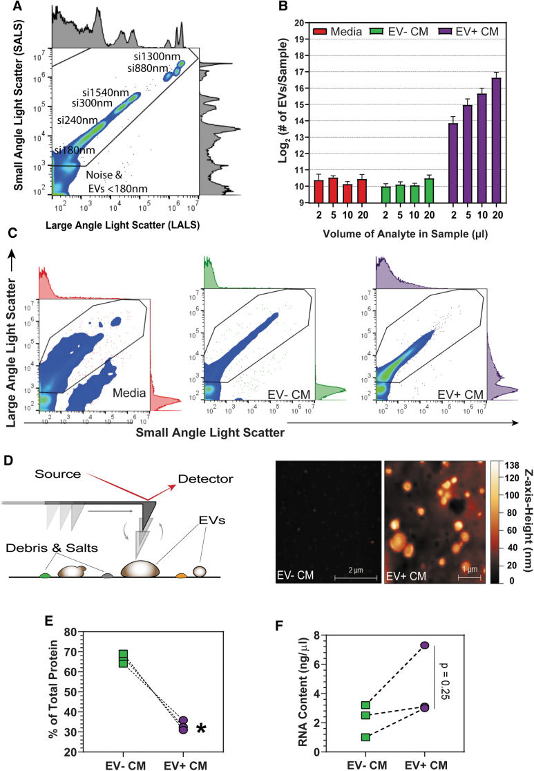
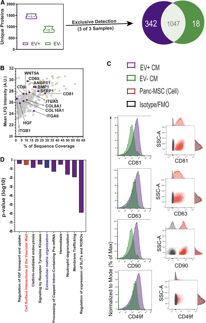
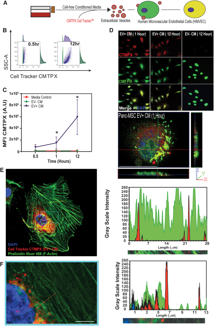
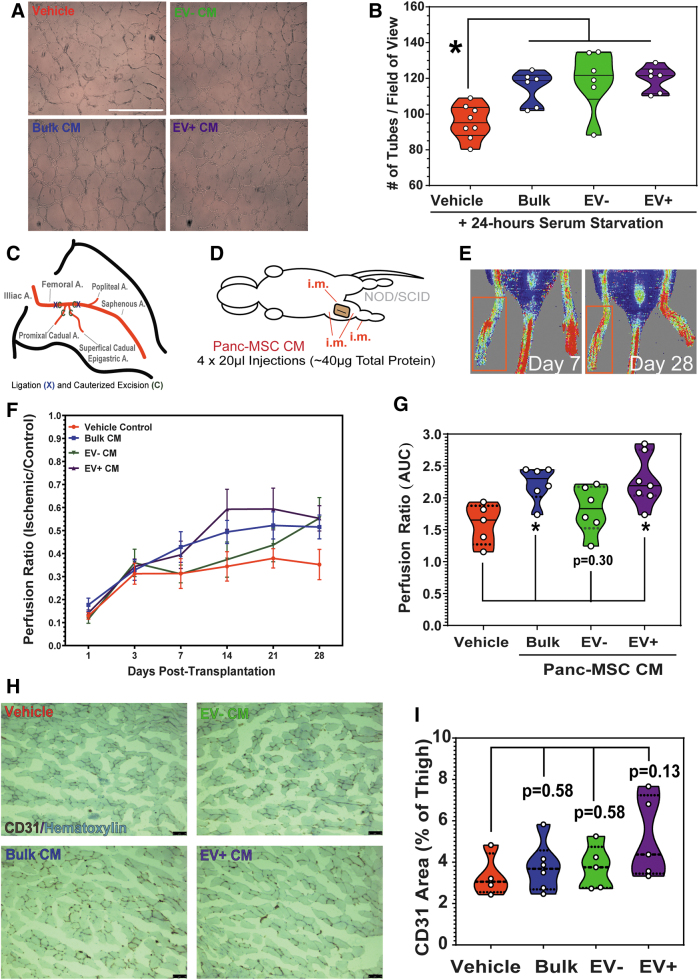
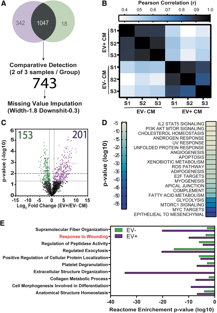
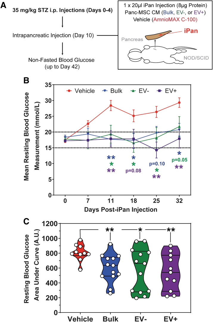
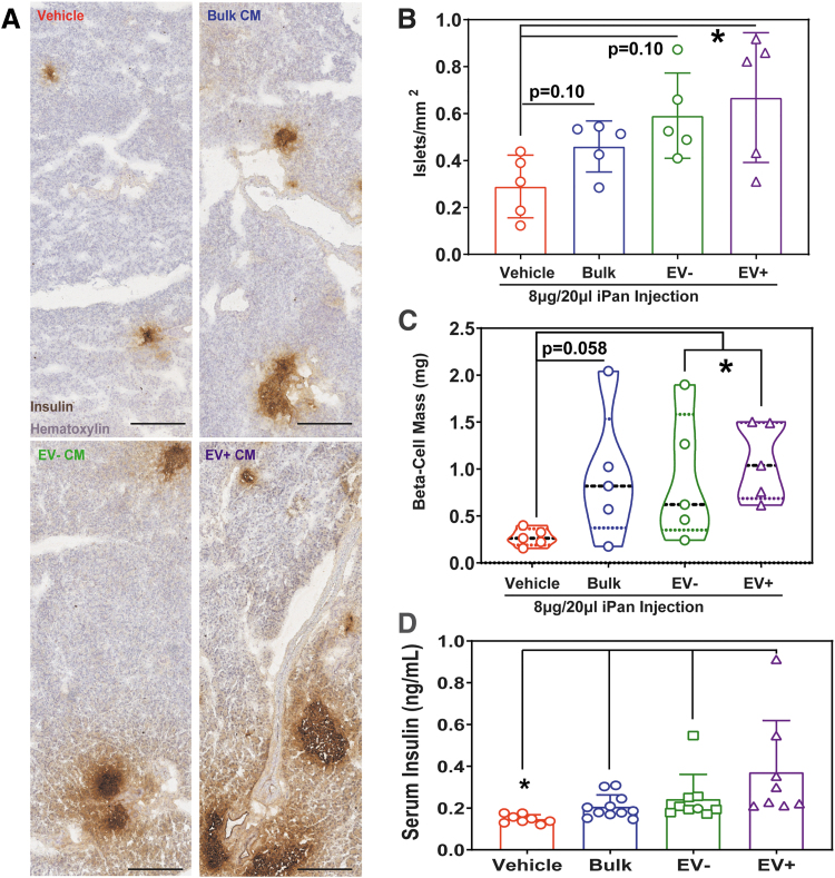
The secretome of mesenchymal stromal cells (MSCs) is enriched for biotherapeutic effectors contained within and independent of extracellular vesicles (EVs) that may support tissue regeneration as an injectable agent. We have demonstrated that the intrapancreatic injection of concentrated conditioned media (CM) produced by bone marrow MSC supports islet regeneration and restored glycemic control in hyperglycemic mice, ultimately providing a platform to elucidate components of the MSC secretome. Herein, we extend these findings using human pancreas-derived MSC (Panc-MSC) as "biofactories" to enrich for tissue regenerative stimuli housed within distinct compartments of the secretome. Specifically, we utilized 100 kDa ultrafiltration as a simple method to debulk protein mass and to enrich for EVs while concentrating the MSC secretome into an injectable volume for preclinical assessments in murine models of blood vessel and islet regeneration. EV enrichment (EV+) was validated using nanoscale flow cytometry and atomic force microscopy, in addition to the detection of classical EV markers CD9, CD81, and CD63 using label-free mass spectrometry. EV+ CM was predominately enriched with mediators of wound healing and epithelial-to-mesenchymal transition that supported functional regeneration in mesenchymal and nonmesenchymal tissues. For example, EV+ CM supported human microvascular endothelial cell tubule formation in vitro and enhanced the recovery of blood perfusion following intramuscular injection in nonobese diabetic/severe combined immunodeficiency mice with unilateral hind limb ischemia. Furthermore, EV+ CM increased islet number and β cell mass, elevated circulating insulin, and improved glycemic control following intrapancreatic injection in streptozotocin-treated mice. Collectively, this study provides foundational evidence that Panc-MSC, readily propagated from the subculture of human islets, may be utilized for regenerative medicine applications.

Keywords: extracellular vesicles; mesenchymal stromal cells; pancreas; proteomics; regenerative medicine; transplantation.


Conflict of interest statement

No competing financial interests exist.

Figures

FIG. 1.
FIG. 1.
EVs are enriched from the secretome of Panc-MSC by 100-kDa ultrafiltration. (A) Representative nanoflow flow cytometry plot of premixed silica calibration beads ranging from 180 to 1,300 nm. si, silica beads. All samples were diluted to 300 μL with 0.22 μm filtered PBS, and 100 μL was acquired using the Apogee A-60 nanoscale flow cytometer. (B) Only EV+ CM produced an increasing linear pattern of EV detection when increasing volumes of samples were analyzed in a fixed acquisition volume. (C) In contrast, the total number of EVs detected in EV− CM did not exceed background detection rates, compared to concentrated media of equal volume. (D) Tapping-mode AFM was used to visualize the 3D architecture of EVs following ultrafiltration. Representative AFM recordings demonstrate that EV+ CM contained vesicle structures with 3D properties consistent of EVs. Notably, parallel structures were not observed in EV− CM. (E) The total mass of protein contained within EV− CM was >EV+ CM. (F) In contrast, EV+ CM demonstrated enriched RNA content compared to EV− CM (*P < 0.05; n = 3). Analyses for significance were determined by Welch's unpaired t-test. EV, extracellular vesicle; MSC, mesenchymal stromal cell; PBS, phosphate-buffered saline; AFM, atomic force microscopy; 3D, three-dimensional; CM, conditioned media; Panc-MSC, pancreas-derived MSC.
FIG. 2.
FIG. 2.
Classical EV membrane markers CD9, CD81, CD63 were exclusively detected in EV+ CM. Despite containing less total protein by mass (Fig. 1E), (A) EV+ CM contained an increased number of unique proteins detected by liquid chromatography–tandem mass spectrometry compared to EV− CM. Three hundred forty-two proteins were exclusively detected within EV+ CM, and 18 proteins were exclusively detected in EV− CM (three out of three samples). (B) Of the 342 exclusive proteins to EV+ CM, classical EV surface markers, including CD9, CD63, and CD81, were detected alongside a number of ribosomal proteins (orange diamonds). Ligands for several biological signaling pathways, such as BMP1, ANGPT1, HGF, and Wnt5a, were exclusively detected in EV+ CM. EV+ CM exclusively contained proteins known to be expressed on the surface of Panc-MSC, such as THY1/CD90 and ITGA6/CD49f. (C) Accordingly, we validated the detection of CD81, CD63, CD90, and CD49f on both the external membrane of EVs in EV+ CM (purple) using nanoscale flow cytometry and on parental cells (red) compared to isotype control (black) using conventional flow cytometry. EV− CM was used as an internal control (green). (D) Proteins exclusively within EV+ CM were associated with several Reactome annotations, including mediators of extracellular matrix organization and/or cellular interactions at the vascular wall. Solid horizontal line indicates mean LFQ intensity (n = 3). LFQ, label-free quantification.
FIG. 3.
FIG. 3.
HMVEC uptake of EVs secreted by human Panc-MSC. (A) Schematic highlighting the cell-independent labeling of Panc-MSC EVs using CMTPX Cell TrackerTM to assess EV uptake in cultured HMVECs labeled with CMFDA (green) Cell Tracker. (B) Representative flow cytometry plots demonstrate that only the EV+ CM fraction was increased. (C) CMTPX fluorescence within cultured HMVEC after 0.5 and 12 h of exposure to EV+ or EV− CM. (D) CMTPX-labeled EVs were detected within adherent CMFDA+ HMVEC at 1 and 12 h, whereas uptake of CMTPX accumulation was not observed in CMFDA+ HMVEC cultured with EV− CM. CMPTX+ EV-like structures were detected within the cytoplasm of HMVEC after 1 h. (EF) CMPTX internalization was observed within 5 min of cultured with EV+ CM. (E) Representative confocal photomicrographs and line scan intensity profiles (yellow line) of CMPTX+ EV-like structures (red) within HMVEC at parallel z-planes with cytoplasmic F-Actin (green). Scale bar = 25 μm. (F) 5 × zoom of selected area (teal) and line scan intensity profile (orange line) of perinuclear and cytoplasmic accumulation of CMTPX fluorescence in HMVEC when cultured with EV+ CM. Scale bar = 10 μm. Data represented as mean ± SEM (*P < 0.05; n = 3). Analyses of significance were performed by Welch's ANOVA with Dunnett's post hoc test assessed at matched timepoints. HMVEC, human microvascular endothelial cell; SEM, standard error of the mean; ANOVA, analysis of variance.
FIG. 4.
FIG. 4.
Panc-MSC CM enhances endothelial function in vitro and in vivo. (A, B) Representative photomicrographs demonstrate enhanced HMVEC tubule formation under serum-starved conditions 24 h after supplementation with ∼20 μg/mL of bulk, EV−, or EV+ CM for 24 h compared to media control. Scale bar = 100 μm. (C) Femoral artery/vein ligation (X) and cauterization (C) were performed to induce unilateral hind limb ischemia in NOD/SCID mice. (D) Mice with unilateral ischemia (perfusion ratios <0.1) received i.m. injections of Panc-MSC bulk, EV−, or EV+ CM (4 × 20 μL injections = ∼40 μg of total protein). To serve as a vehicle control, equivalent volumes of basal AmnioMAX were injected. (E) Perfusion ratios were quantified by LDPI intensities between ischemic and nonischemic limbs up to day 28. Orange squares indicate area of foot measured. (F) LDPI up to 28 days postinjection demonstrates enhanced recovery of blood perfusion following the intramuscular injection of bulk or EV+ CM, as determined by (G) AUC. EV− CM injection was statistically comparable to vehicle control. (H) Representative photomicrographs of ischemic thigh muscle tissues following 28-days postinjection with vehicle control, bulk, EV−, or EV+ CM. (I) A trending increase of CD31+ area was observed in thigh muscles injected with EV+ CM, although statistically comparable to muscle tissue injected with vehicle control. Scale bar = 100 μm. Data represented as mean ± SEM (*P < 0.05; n = 5–7, N = 3). Analyses of significance were determined by Welch's ANOVA with Dunnett's post hoc test. NOD/SCID, nonobese diabetic/severe combined immunodeficiency; i.m., intramuscular; LDPI, laser doppler perfusion imaging; AUC, area under the curve.
FIG. 5.
FIG. 5.
The secretome of Panc-MSC was enriched with mediators of epithelial to mesenchymal transition and response to wound healing annotations. (A) Workflow for the analyses of proteins common to both EV− and EV+ subfractions. Of the total proteins identified, 1,047 proteins were detected in at least one out of three EV− or EV+ samples, and 743 of 1,047 were detected in two of three samples within EV− and EV+ CM. Missing value imputation was performed with width and downshift set to 1.8 and 0.3 in Perseus software, respectively. (B) Despite common detection, a unique signature of EV− and EV+ CM was identified, and the reproducibility of the EV− and EV+ CM production was validated by Pearson correlation analysis. (C) One hundred fifty-three and 201 common proteins were commonly detected at statistically distinct levels within EV− (green dots) and EV+ (purple dots) CM, respectively. (D) Statistically comparable proteins (black dots) were associated with the Reactome annotations for angiogenesis and effectors of epithelial to mesenchymal transition. (E) Statistically distinct proteins were associated with the Reactome annotations for supramolecular fiber organization and responses to wounding.
FIG. 6.
FIG. 6.
Intrapancreatic injection of Panc-MSC EV+ or EV− CM stabilized blood glucose levels in STZ-treated hyperglycemic mice. (A) β Cell ablation was induced in NOD/SCID mice through intraperitoneal injection of STZ (35 mg/kg/day) on days 0–4. At day 10, hyperglycemic (nonfasted blood glucose measurements between 14.5 and 24.5 mmol/L) mice were transplanted in a blinded manner by iPan injection of concentrated AmnioMAX C-100 basal media or concentrated bulk EV− or EV+ CM containing ∼8 μg total protein. Resting blood glucose was measured weekly for up to day 42 (32 days postinjection). (B) Mean nonfasted resting blood glucose demonstrated a significant or trending decrease of hyperglycemia in mice receiving EV−, EV+, or bulk CM. Horizontal dotted lines represent upper and lower glycemia levels in mice used for iPan transplantation studies at day 10. (C) iPan injection of bulk, EV−, or EV+ CM generated by Panc-MSC significantly reduced and stabilized hyperglycemia, as determined by AUC. (B) Analyses of significance were determined by repeated measures two-way ANOVA with post hoc Tukey's t-test compared to vehicle control. (C) Analyses of significance (*P < 0.05, **P < 0.01) were determined by Welch's ANOVA with Dunnett's post hoc test. STZ, streptozotocin; iPan, intrapancreatic.
FIG. 7.
FIG. 7.
Intrapancreatic injection of Panc-MSC EV+ CM increased islet number, β cell mass, and serum insulin in STZ-treated hyperglycemic mice. (A) Representative photomicrographs of immunohistochemical detection of insulin-containing islets in pancreas cryosections separated by >150 μm. (B) EV+ CM significantly increased the number of islets/mm2 after 32 days, compared to mice injected with vehicle control. (C) As a result, EV+ CM injection significantly increased β cell mass. Although not significant, β cell mass was elevated in the pancreas of mice which received an intrapancreatic injection of bulk and EV− CM. Data as mean ± SEM (n = 5). (D) Elevated insulin was detected in blood serum of mice injected with bulk, EV−, or EV+ CM. Data represented as mean ± SEM (n = 10–13, N = 3). Analyses of significance (*P < 0.05) were determined by Welch's ANOVA with Dunnett's post hoc test.

Similar articles

Cited by

References

    1. Caplan AI and Correa D. (2011). The MSC: an injury drugstore. Cell Stem Cell 9:11–15. - PMC - PubMed
    1. Caplan AI. (2016). MSCs: the sentinel and safe-guards of injury. J Cell Physiol 231:1413–1416. - PubMed
    1. Squillaro T, Peluso G and Galderisi U. (2016). Clinical trials with mesenchymal stem cells: an update. Cell Transplant 25:829–848. - PubMed
    1. Bell GI, Broughton HC, Levac KD, Allan DA, Xenocostas A and Hess DA. (2011). Transplanted human bone marrow progenitor subtypes stimulate endogenous islet regeneration and revascularization. Stem Cells Dev 21:97–109. - PubMed
    1. Leibacher J and Henschler R. (2016). Biodistribution, migration and homing of systemically applied mesenchymal stem/stromal cells. Stem Cell Res Ther 7:7. - PMC - PubMed

Publication types

MeSH terms

LinkOut - more resources