. 2021 Oct;17(10):3160-3174.
doi: 10.1080/15548627.2020.1871211. Epub 2021 Jan 25.

Autophagy inhibition rescues structural and functional defects caused by the loss of mitochondrial chaperone Hsc70-5 in Drosophila

Affiliations

Autophagy inhibition rescues structural and functional defects caused by the loss of mitochondrial chaperone Hsc70-5 in Drosophila

Jun-Yi Zhu et al. Autophagy. 2021 Oct.

Abstract

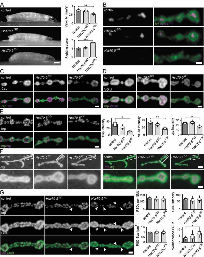
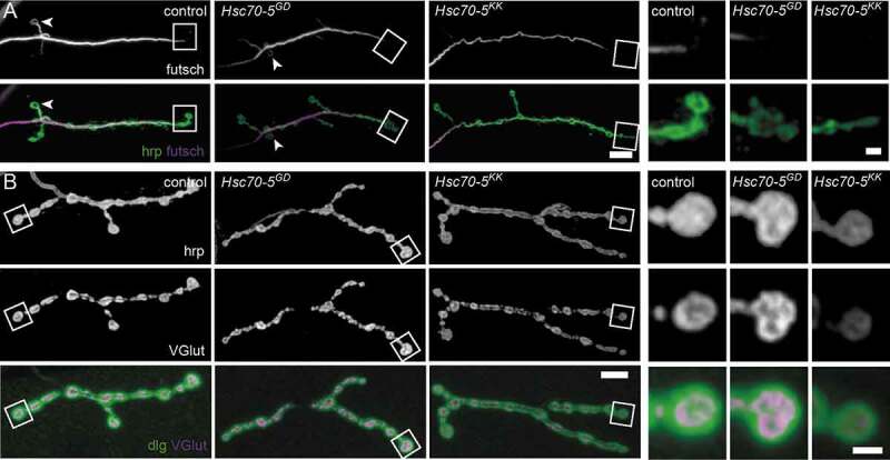
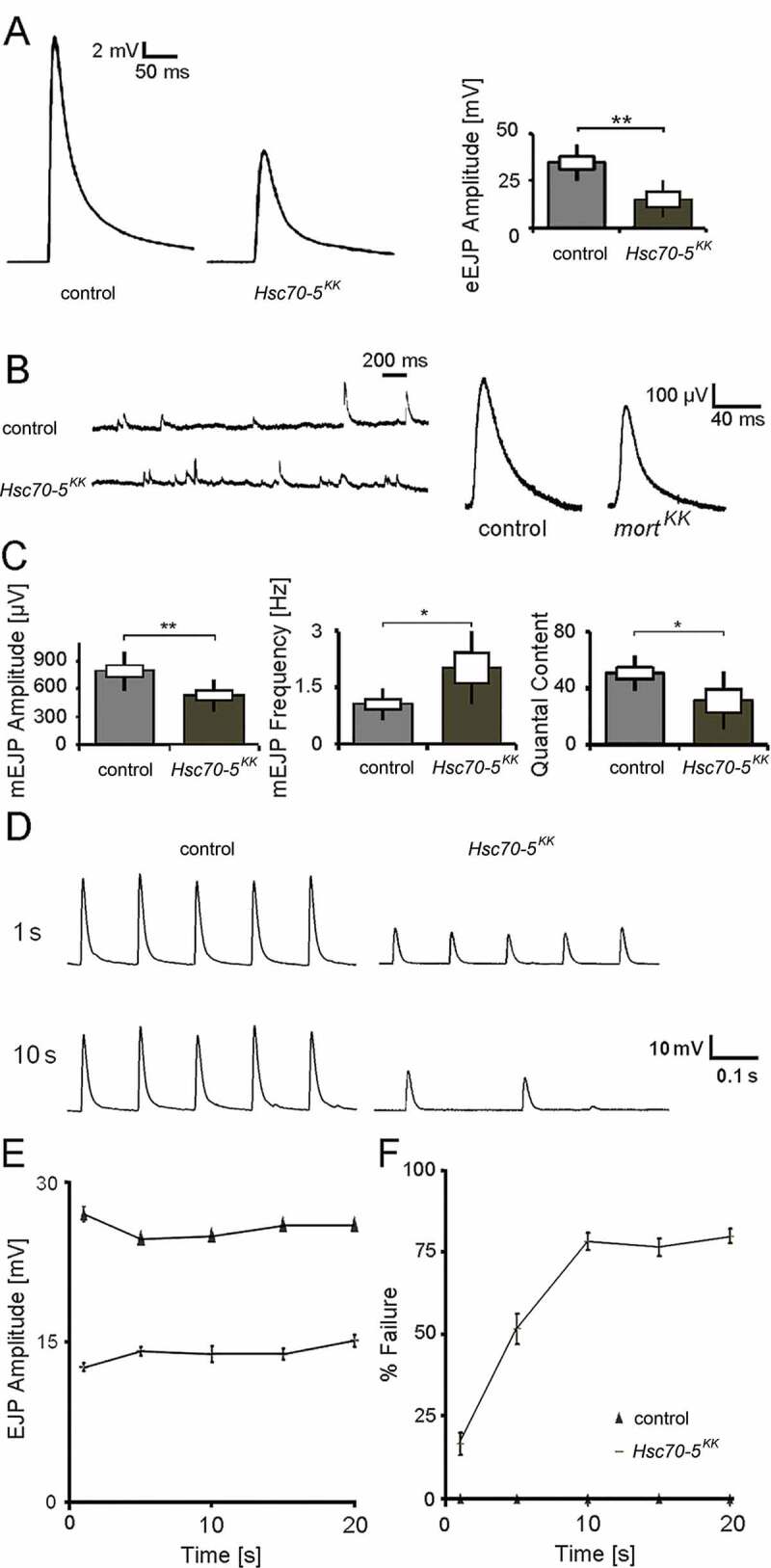
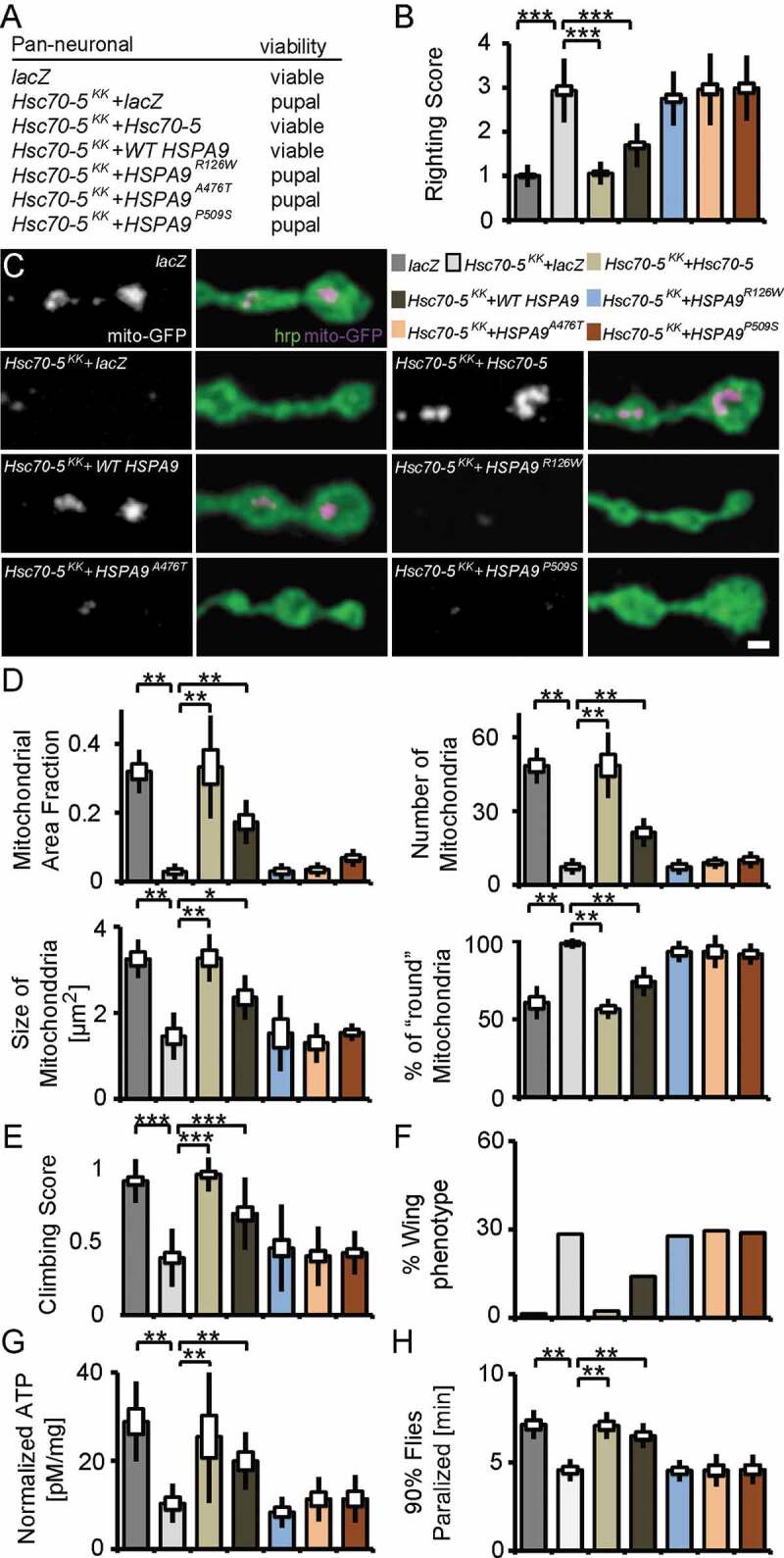
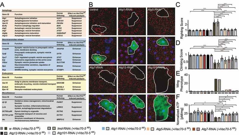
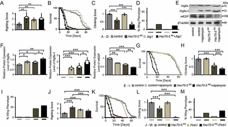
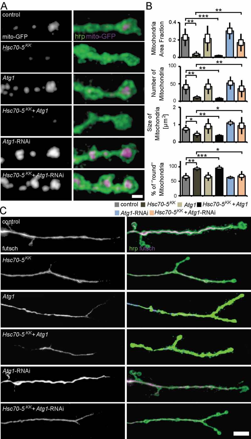
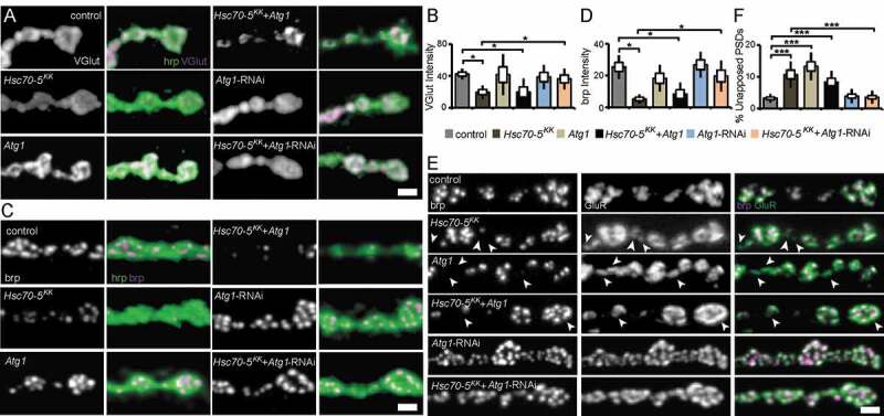
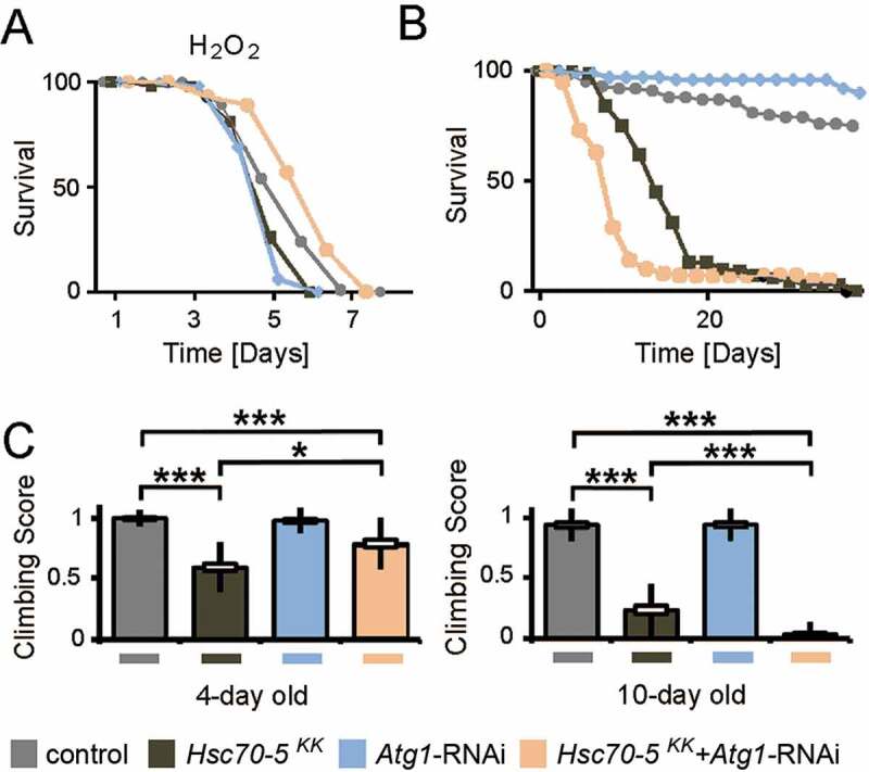
We investigated in larval and adult Drosophila models whether loss of the mitochondrial chaperone Hsc70-5 is sufficient to cause pathological alterations commonly observed in Parkinson disease. At affected larval neuromuscular junctions, no effects on terminal size, bouton size or number, synapse size, or number were observed, suggesting that we studied an early stage of pathogenesis. At this stage, we noted a loss of synaptic vesicle proteins and active zone components, delayed synapse maturation, reduced evoked and spontaneous excitatory junctional potentials, increased synaptic fatigue, and cytoskeleton rearrangements. The adult model displayed ATP depletion, altered body posture, and susceptibility to heat-induced paralysis. Adult phenotypes could be suppressed by knockdown of dj-1β, Lrrk, DCTN2-p50, DCTN1-p150, Atg1, Atg101, Atg5, Atg7, and Atg12. The knockdown of components of the macroautophagy/autophagy machinery or overexpression of human HSPA9 broadly rescued larval and adult phenotypes, while disease-associated HSPA9 variants did not. Overexpression of Pink1 or promotion of autophagy exacerbated defects.Abbreviations: AEL: after egg laying; AZ: active zone; brp: bruchpilot; Csp: cysteine string protein; dlg: discs large; eEJPs: evoked excitatory junctional potentials; GluR: glutamate receptor; H2O2: hydrogen peroxide; mEJP: miniature excitatory junctional potentials; MT: microtubule; NMJ: neuromuscular junction; PD: Parkinson disease; Pink1: PTEN-induced putative kinase 1; PSD: postsynaptic density; SSR: subsynaptic reticulum; SV: synaptic vesicle; VGlut: vesicular glutamate transporter.

Keywords: Atg1; Hsc70-5; microtubule; mitochondria; mitophagy; rapamycin; synapse.


Conflict of interest statement

All authors declare no conflict of interest.

Figures

Figure 1.
Figure 1.
Characterization of the neuromuscular junction in presymptomatic and symptomatic larval. (A) Average larval crawling velocity and righting reflex at L3 stage. Scale bar: 0.25 mm. (B) Confocal images of larval NMJ expressing mito-GFP (magenta) and labeled with hrp (green). Scale bar: 2 μm. (C-E) Representative synaptic boutons from NMJs of control, elav>Hsc70-5GD13957 and elav>Hsc70-5KK100233 larvae. Anti-hrp staining (C-E, green) was used to visualize synaptic membranes. Synaptic vesicle proteins Csp (C) and VGlut (D) and active zone component brp (E) were shown in magenta. Scale bar: 2 μm. (F) Confocal images of larval NMJ labeled with hrp (magenta) and Drosophila dlg (green) used to visualize the sub-synaptic reticulum (SSR). Scale bar: 5 μm, enlargement: 2 μm. (G) Confocal images of larval NMJ labeled with brp (magenta) and GluR (green) were used to visualize PSDs (postsynaptic densities). Arrowheads pointing out presynaptic brp labels were not detected in PSDs. Scale bar: 2 μm. The standard error of mean and standard deviation are shown as a box and a black line. * p < 0.05, ** p < 0.01, *** p < 0.001
Figure 2.
Figure 2.
Loss of Hsc70-5 function is associated with MT cytoskeleton defects. (A) Confocal images of larval NMJ labeled with hrp (green) and futsch (MT cytoskeleton; magenta). Scale bar: 5 μm, enlargement: 2 μm. Arrowheads pointing at NMJ loops. (B) Confocal images of larval NMJ labeled with hrp (gray), VGlut (gray and magenta), and dlg (green). Scale bar: 10 μm, enlargement: 2 μm
Figure 3.
Figure 3.
Electrophysiological characterization of symptomatic larvae. (A) Representative traces of eEJPs from control and elav>Hsc70-5KK100233 larvae and quantification of eEJP amplitudes. Stimulation artifacts were removed from eEJP traces. (B) Representative mEJP recording in control and elav>Hsc70-5KK100233 larvae. Quantification of (C) mEJP amplitude, mEJP frequency and quantal content in control and elav>Hsc70-5KK100233 larvae. (D) Representative traces from 10 Hz stimulation displaying five successive synaptic responses from each genotype after 1 and 10 s stimulation. Stimulation artifacts were removed for demonstration. (E) Quantification of eEJP amplitude in response to 10 Hz stimulation in control and elav>Hsc70-5KK100233 larvae. (F) Quantification of % failure of eEJP in response to 10 Hz stimulation in control and elav>Hsc70-5KK100233 larvae. The standard error of mean and standard deviation are shown as a box and a black line. * p < 0.05, ** p < 0.01
Figure 4.
Figure 4.
Drosophila Hsc70-5 and human WT HSPA9 but not disease variants rescued Hsc70-5 knockdown phenotypes. (A) Hsc70-5 knockdown in combination with lacZ overexpression to control Gal4 dilution at 25°C caused pupal lethality. Overexpression of Drosophila Hsc70-5 and human WT HSPA9 in the Hsc70-5KK100233 background unlike HSPA9R126W, HSPA9A476T and HSPA9P509S variants rescued pupal lethality. (B) Average larval righting reflex at L3 stage with overexpression of Hsc70-5, WT HSPA9, HSPA9R126W, HSPA9A476T and HSPA9P509S in the elav>Hsc70-5KK100233 background. (C) Confocal images of larval NMJ labeled with hrp (green) and mito-GFP (magenta), and (D) quantification of mitochondrial parameters. Scale bar: 2 μm. (E) Climbing ability of 4-d-old flies, (F) Percentage of flies with defective wing phenotype, and (G) ATP levels in fly heads after expressing Hsc70-5, WT HSPA9, HSPA9R126W, HSPA9A476T and HSPA9P509S in the elav>Hsc70-5KK100233 background. (H) Hsc70-5 knockdown accelerated heat-shock induced paralysis in flies at 39.5°C. The co-overexpression of Hsc70-5 and WT HSPA9 unlike HSPA9R126W, HSPA9A476T and HSPA9P509S rescued this defect. The standard error of mean and standard deviation are shown as a box and a black line. * p < 0.05, ** p < 0.01, *** p < 0.001
Figure 5.
Figure 5.
A genetic screen identified autophagy-related proteins as modifiers of Hsc70-5 knockdown phenotypes. (A) A genetic screen identified several modifiers that modified Hsc70-5 knockdown-induced phenotypes. (B) Increase in mCherry.Atg8a puncta in the larval fat body following starvation. There was no difference in the expression of mCherry.Atg8a between GFP +ve and -ve cells in control overexpressing lacZ (in GFP +ve cell). Expression of RNAi against autophagy-related genes (Atg1, Atg101 Atg5, Atg7, and Atg12) inhibited starvation-induced autophagy in GFP +ve cells compared to cells in the vicinity. Scale bar: 15 μm. (C) Righting reflex in larvae, (D) Climbing ability of 4-d-old flies, (E) Percentage of flies with defective wing phenotype, and (F) ATP levels in fly heads after pan-neuronal knockdown of Atg1, Atg101 Atg5, Atg7, and Atg12 in control and the elav>Hsc70-5KK100233,tub-GAL80ts background. The standard error of mean and standard deviation are shown as a box and a black line. * p < 0.05, *** p < 0.001
Figure 6.
Figure 6.
Autophagy induction did not rescue symptomatic phenotypes in Hsc70-5 knockdown flies. (A) Quantification of larval righting reflex, (B) Lifespan of flies at 25°C, (C) Climbing ability of 4-d-old flies and (D) Percentage of flies with defective wing phenotype expressing Atg1 in the elav>Hsc70-5KK100233,tub-GAL80ts background. (E) Western blot showing levels of Atg8a, Atg8a-II, and ref(2)P in rapamycin-treated flies in the elav>Hsc70-5KK100233,tub-GAL80ts background. βTub56D was used as a control. (F) Quantifications of protein levels of Atg8a, Atg8a-II, and ref(2)P. (G) The lifespan of flies with rapamycin treatment in the elav>Hsc70-5KK100233,tub-GAL80ts background at 25°C. (H) The climbing ability of 4-d-old flies, and (I) percentage of flies with defective wing phenotype with rapamycin treatment in the elav>Hsc70-5KK100233,tub-GAL80ts background. (J) Quantification of larval righting reflex, (K) Lifespan of flies at 25°C, (L) Climbing ability of 4-d-old flies, and (M) percentage of flies with defective wing phenotype expressing Pink1 in the elav>Hsc70-5KK100233,tub-GAL80ts background. The standard error of mean and standard deviation are shown as a box and a black line. * p < 0.05, ** p < 0.01, *** p < 0.001
Figure 7.
Figure 7.
Autophagy inhibition alleviated mitochondrial and MT defects caused by Hsc70-5 knockdown. (A) Confocal images of larval NMJ expressing mito-GFP (magenta) labeled with hrp (green). Scale bar: 2 μm. (B) Quantification of mitochondrial area fraction, number, size, and morphology. (C) Confocal images of NMJ labeled with hrp (green) and futsch (magenta). Scale bar: 10 μm. The standard error of mean and standard deviation are shown as a box and a black line. * p < 0.05, ** p < 0.01, *** p < 0.001
Figure 8.
Figure 8.
Autophagy inhibition alleviated synaptic defects caused by Hsc70-5 knockdown. (A and C) Confocal images of larval NMJ labeled with hrp (green), VGlut, and brp (magenta). Scale bar: 2 μm. (B and D) Quantification of VGlut and brp level at NMJ following Atg1 overexpression and knockdown in elav>Hsc70-5KK100233 background. (E) Confocal images of larval NMJ labeled with brp (magenta) and GluR (green). Arrowheads pointing out regions where presynaptic brp labels were not detected in PSDs. Scale bar: 2 μm (F) Quantification of unapposed glutamate receptor fields. The standard error of mean and standard deviation are shown as a box and a black line. * p < 0.05, *** p < 0.001
Figure 9.
Figure 9.
Concomitant Atg1 and Hsc70-5 knockdown rescued longevity under oxidative stress but was detrimental upon aging in baseline conditions. (A) Lifespan of flies at 25°C following induction of generalized oxidative stress. Flies were fed with 5% hydrogen peroxide sucrose solution. (B) Lifespan of flies at 25°C on the standard diet. (C) The climbing ability of 4 – and 10-d-old flies expressing Atg1 knockdown in the elav>Hsc70-5KK100233,tub-GAL80ts background. The standard error of mean and standard deviation are shown as a box and a black line. * p < 0.05, *** p < 0.001

References

    1. Dauer W, Przedborski S.. Parkinson’s disease: mechanisms and models. Neuron. 2003. Sep 11;39(6):889–909. - PubMed
    1. Picconi B, Piccoli G, Calabresi P. Synaptic dysfunction in Parkinson’s disease. Adv Exp Med Biol. 2012;970:553–572. - PubMed
    1. Bras J, Guerreiro R, SnapShot: HJ. Genetics of Parkinson’s disease. Cell. 2015. Jan 29;160(3):570–570 e1. - PubMed
    1. Miura E, Hasegawa T, Konno M, et al. VPS35 dysfunction impairs lysosomal degradation of alpha-synuclein and exacerbates neurotoxicity in a Drosophila model of Parkinson’s disease. Neurobiol Dis. 2014. Nov;71:1–13. - PubMed
    1. Misgeld T, Schwarz TL. Mitostasis in neurons: maintaining mitochondria in an extended cellular architecture. Neuron. 2017. Nov 1;96(3):651–666. - PMC - PubMed

Publication types

MeSH terms

LinkOut - more resources