

Perioperative abnormal electroencephalography in a later-stage elderly with septic shock: a case report
- PMID: 33404769
- PMCID: PMC7786879
- DOI: 10.1186/s40981-020-00409-5
Perioperative abnormal electroencephalography in a later-stage elderly with septic shock: a case report
Abstract
Background: Patients with sepsis often exhibit abnormal patterns of electroencephalogram (EEG). We report an abnormal EEG pattern in a later-stage elderly patient with septic shock and EEG analysis results.
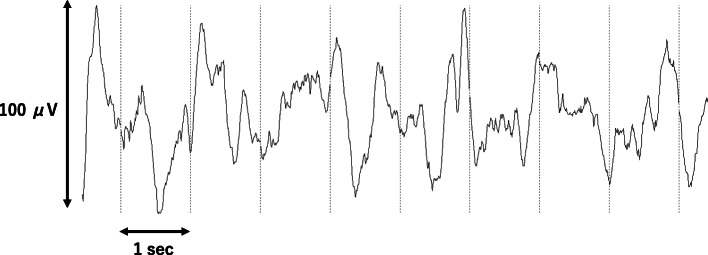
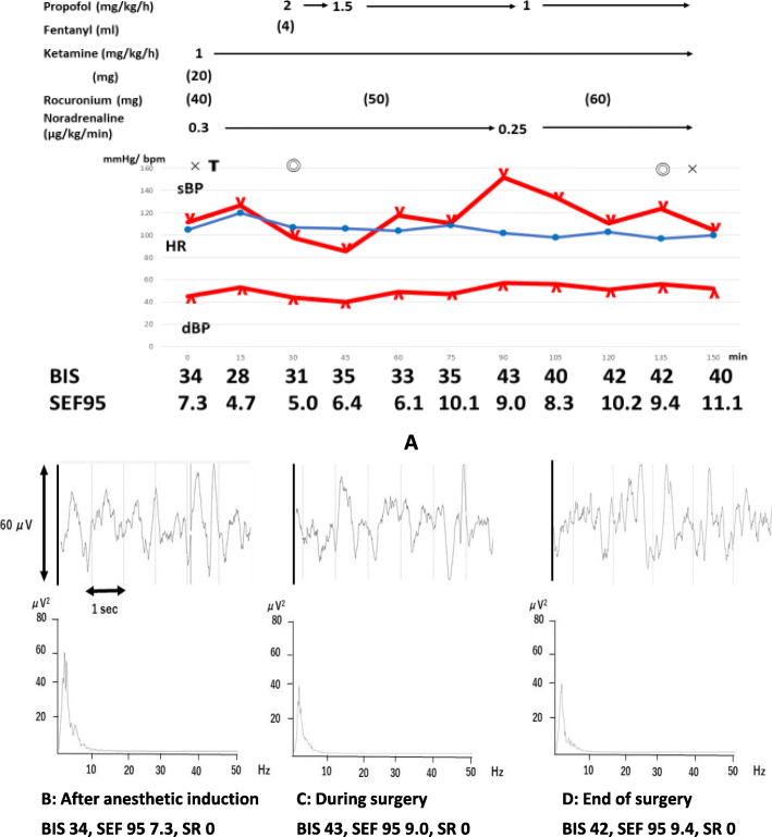
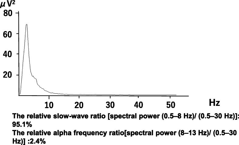
Case presentation: An 88-year-old woman with bowel perforation underwent emergency Hartmann surgery. On admission to the operating room, she exhibited septic shock. Her bispectral index value was 30 before anesthesia induction, and the EEG displayed slow waves without burst and suppression throughout the surgery. The relative slow-wave ratio [spectral power (0.5-8 Hz)/(0.5-30 Hz)] from anesthetic induction to the end of surgery was 95.1%, whereas the relative alpha frequency [spectral power (8-13 Hz)/(0.5-30 Hz)] was only 2.4%. Although without preoperative neurological abnormalities, she developed postoperative delirium after admission to the intensive care unit.
Conclusions: Intraoperative continuous EEG monitoring in elderly patients with sepsis may be useful to predict sepsis-associated encephalopathy. Therefore, continuous EEG monitoring may improve neurological outcomes.
Keywords: Electroencephalogram; Ketamine; Postoperative delirium; Sepsis; Sepsis-associated encephalopathy.
Conflict of interest statement
The authors declare that they have no conflict of interest.
Figures
References
-
- Ma Y, Ouyang B, Guan X. Use of quantitative electroencephalogram in patients with septic shock. Zhonghua Yi Xue Za Zhi. 2016;96:195–198. - PubMed
-
- van Dellen E, van Dellen E, van der Kooi AW, Numan T, Koek HL, Klijn FA, Buijsrogge MP, et al. Decreased functional connectivity and disturbed directionality of information flow in the electroencephalography of intensive care unit patients with delirium after cardiac surgery. Anesthesiology. 2014;121:328–335. doi: 10.1097/ALN.0000000000000329. - DOI - PubMed
LinkOut - more resources
Full Text Sources
Other Literature Sources