

Chest radiograph features of multisystem inflammatory syndrome in children (MIS-C) compared to pediatric COVID-19
- PMID: 33404786
- PMCID: PMC7785920
- DOI: 10.1007/s00247-020-04921-9
Chest radiograph features of multisystem inflammatory syndrome in children (MIS-C) compared to pediatric COVID-19
Abstract
Background: Although the radiographic features of coronavirus disease 2019 (COVID-19) in children have been described, the distinguishing features of multisystem inflammatory syndrome in children (MIS-C) associated with COVID-19 are not well characterized.
Objective: We compared the chest radiographic findings of MIS-C with those of COVID-19 and described other distinguishing imaging features of MIS-C.
Materials and methods: We performed a retrospective case series review of children ages 0 to 18 years who were hospitalized at Children's Healthcare of Atlanta from March to May 2020 and who either met the Centers for Disease Control and Prevention (CDC) case definition for MIS-C (n=11) or who had symptomatic, laboratory-confirmed COVID-19 (n=16). Two radiologists reviewed the most severe chest radiographs for each patient. The type and distribution of pulmonary opacities and presence or absence of pleural effusions were recorded. The chest radiographs were categorized based on potential COVID-19 imaging findings as typical, indeterminate, atypical or negative. An imaging severity score was also assigned using a simplified version of the Radiographic Assessment of Lung Edema Score. Findings were statistically compared between patients with MIS-C and those with COVID-19. Additional imaging findings of MIS-C were also described.
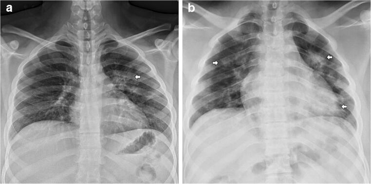
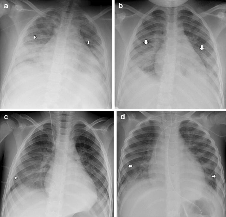
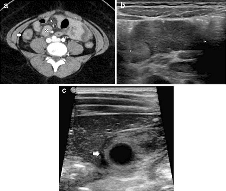
Results: Radiographic features of MIS-C included pleural effusions (82% [9/11]), pulmonary consolidations (73% [8/11]) and ground glass opacities (91% [10/11]). All of the lung opacities (100% [10/10]) were bilateral, and the majority of the pleural effusions (67% [6/9]) were bilateral. Compared to children with COVID-19, children with MIS-C were significantly more likely to develop pleural effusions on chest radiograph (82% [9/11] vs. 0% [0/0], P-value <0.01) and a lower zone predominance of pulmonary opacifications (100% [10/10] vs. 38% [5/13], P-value <0.01). Children with MIS-C who also had abdominal imaging had intra-abdominal inflammatory changes.
Conclusion: Key chest radiographic features of MIS-C versus those of COVID-19 were pleural effusions and lower zone pulmonary opacifications as well as intra-abdominal inflammation. Elucidating the distinguishing radiographic features of MIS-C may help refine the case definition and expedite diagnosis and treatment.
Keywords: Abdomen; Chest; Children; Computed tomography; Coronavirus disease 2019; Multisystem inflammatory syndrome in children; Radiography; Ultrasound.
Conflict of interest statement
Christina A. Rostad has received royalties unrelated to this manuscript to Emory University from Meissa Vaccines Inc. She has also received funds to her institution to conduct clinical research unrelated to this manuscript from MedImmune, Regeneron, Paxvax, Pfizer, GSK, Merck, Novavax, Sanofi-Pasteur and Micron.
Figures
References
-
- World Health Organization (2020) Coronavirus disease 2019 (COVID-19) Situation Report — 51. https://www.who.int/docs/default-source/coronaviruse/situation-reports/2.... Accessed 4 July 2020
Publication types
MeSH terms
Supplementary concepts
LinkOut - more resources
Full Text Sources
Other Literature Sources
Medical
Miscellaneous
