Review
. 2021 Jan;383(1):457-472.
doi: 10.1007/s00441-020-03348-w. Epub 2021 Jan 6.

Topographic organization in the olfactory bulb

Affiliations
Review

Topographic organization in the olfactory bulb

Claudia Lodovichi. Cell Tissue Res. 2021 Jan.

Abstract

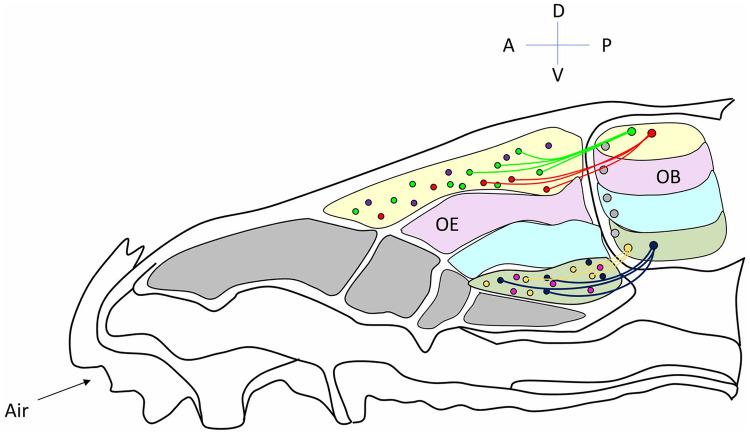
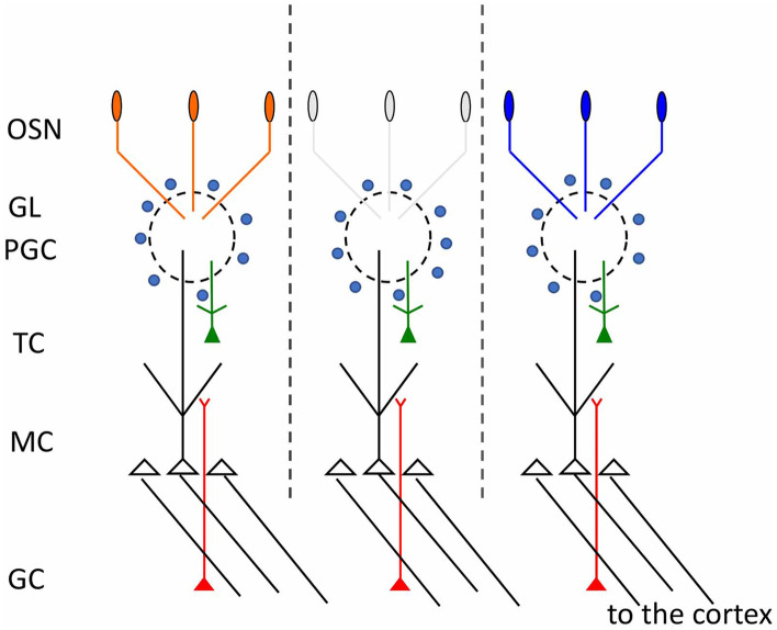
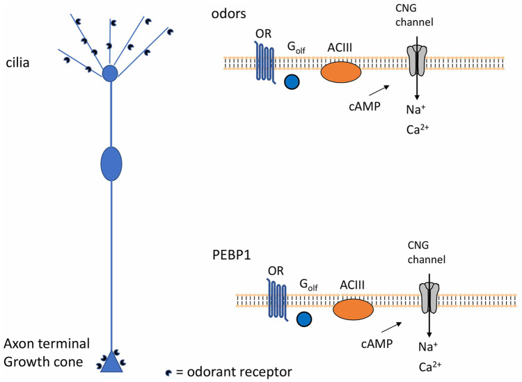
The ability of the olfactory system to detect and discriminate a broad spectrum of odor molecules with extraordinary sensitivity relies on a wide range of odorant receptors and on the distinct architecture of neuronal circuits in olfactory brain areas. More than 1000 odorant receptors, distributed almost randomly in the olfactory epithelium, are plotted out in two mirror-symmetric maps of glomeruli in the olfactory bulb, the first relay station of the olfactory system. How does such a precise spatial arrangement of glomeruli emerge from a random distribution of receptor neurons? Remarkably, the identity of odorant receptors defines not only the molecular receptive range of sensory neurons but also their glomerular target. Despite their key role, odorant receptors are not the only determinant, since the specificity of neuronal connections emerges from a complex interplay between several molecular cues and electrical activity. This review provides an overview of the mechanisms underlying olfactory circuit formation. In particular, recent findings on the role of odorant receptors in regulating axon targeting and of spontaneous activity in the development and maintenance of synaptic connections are discussed.

Keywords: Electrical activity; Neuronal circuits; Odorant receptor; Olfactory bulb; Topographic map.


Conflict of interest statement

The author declares that there is no conflict of interest.

Figures

Fig. 1
Fig. 1
From the olfactory epithelium to the olfactory bulb. Schematic of the subdivision of the olfactory epithelium (OE) in zones along the dorso-ventral axis and the corresponding zones in the olfactory bulb (OB). Areas of different colors indicate the different zones. In the OE, within each zone, olfactory sensory neurons (OSNs) expressing different odorant receptors (ORs), indicated by circles in different colors, are almost randomly distributed. OSNs expressing the same OR converge to form glomeruli in specific locations of the OB. The distribution of OSNs in zones along the dorso-ventral axis of the OE is reflected in the location of the related glomeruli in corresponding zones along the dorso-ventral axis of the OB. Indeed, ONSs located in the most dorsal zone of the OE, project and converge to form glomeruli in the most dorsal area of the bulb. OSNs located in the most ventral area of the OE, project to the most ventral part of the  OB. OSNs which occupy intermediate positions in the OE, project in corresponding zones in the OB, along the dorso-ventral axis. For simplicity, only projections of OSNs located in the most dorsal and ventral areas, respectively, are shown. D dorsal, V ventral, A anterior, P posterior
Fig. 2
Fig. 2
Odor columns in the olfactory bulb. Schematic diagram of the connectivity between pre- and postsynaptic cells in the olfactory bulb. Each glomerulus defines a functional unit, indicated also as odor column (depicted between the dashed vertical lines). OSN olfactory sensory neurons, GL glomerular layer, PGC periglomerular cells, TC tufted cells, MC mitral cells, GC granule cells
Fig. 3
Fig. 3
Link between homologous glomeruli. Schematic diagram of the intrabulbar link between homologous glomeruli. OSN olfactory sensory neurons, GL glomerular layer, ETC external tufted cells, EPL external plexiform layer, MC mitral cells, MCL mitral cell layer, IPL internal plexiform layer, GC granule cells, GCL granule cell layer
Fig. 4
Fig. 4
Localized cAMP and Ca2+ synthesis at the cilia and at the axon terminal of olfactory sensory neurons (OSNs). Odorant receptors are expressed in specific locations in OSNs: at the cilia and at the axon terminal. In both sites (i.e., cilia and axon terminal) ORs are coupled to localized increases of cAMP and Ca2+. OR odorant receptor, ACIII adenylyl cyclase III, CNG channels cyclic nucleotide gated channels

References

    1. Akiyama H, Fukuda T, Tojima T, Nikolaev VO, Kamiguchi H. Cyclic nucleotide control of microtubule dynamics for axon guidance. The Journal of neuroscience: The official journal of the Society for Neuroscience. 2016;36:5636–5649. - PMC - PubMed
    1. Assens A, Dal Col JA, Njoku A, Dietschi Q, Kan C, Feinstein P, Carleton A, Rodriguez I. Alteration of Nrp1 signaling at different stages of olfactory neuron maturation promotes glomerular shifts along distinct axes in the olfactory bulb. Development. 2016;143:3817–3825. - PMC - PubMed
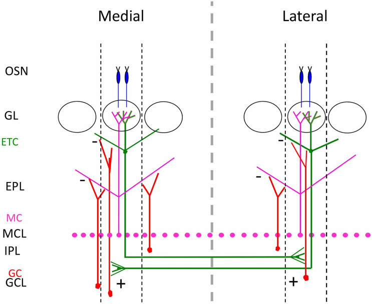
    1. Aungst JL, Heyward PM, Puche AC, Karnup SV, Hayar A, Szabo G, Shipley MT. Centre-surround inhibition among olfactory bulb glomeruli. Nature. 2003;426:623–629. - PubMed
    1. Barnea G, O'Donnell S, Mancia F, Sun X, Nemes A, Mendelsohn M, Axel R. Odorant receptors on axon termini in the brain. Science. 2004;304:1468. - PubMed
    1. Belluscio L, Gold GH, Nemes A, Axel R. Mice deficient in G(olf) are anosmic. Neuron. 1998;20:69–81. - PubMed

LinkOut - more resources