. 2021 Jul 8:44:49-67.
doi: 10.1146/annurev-neuro-101920-112225. Epub 2021 Jan 6.

Astrocytes and Behavior

Affiliations

Astrocytes and Behavior

Paulo Kofuji et al. Annu Rev Neurosci. .

Abstract

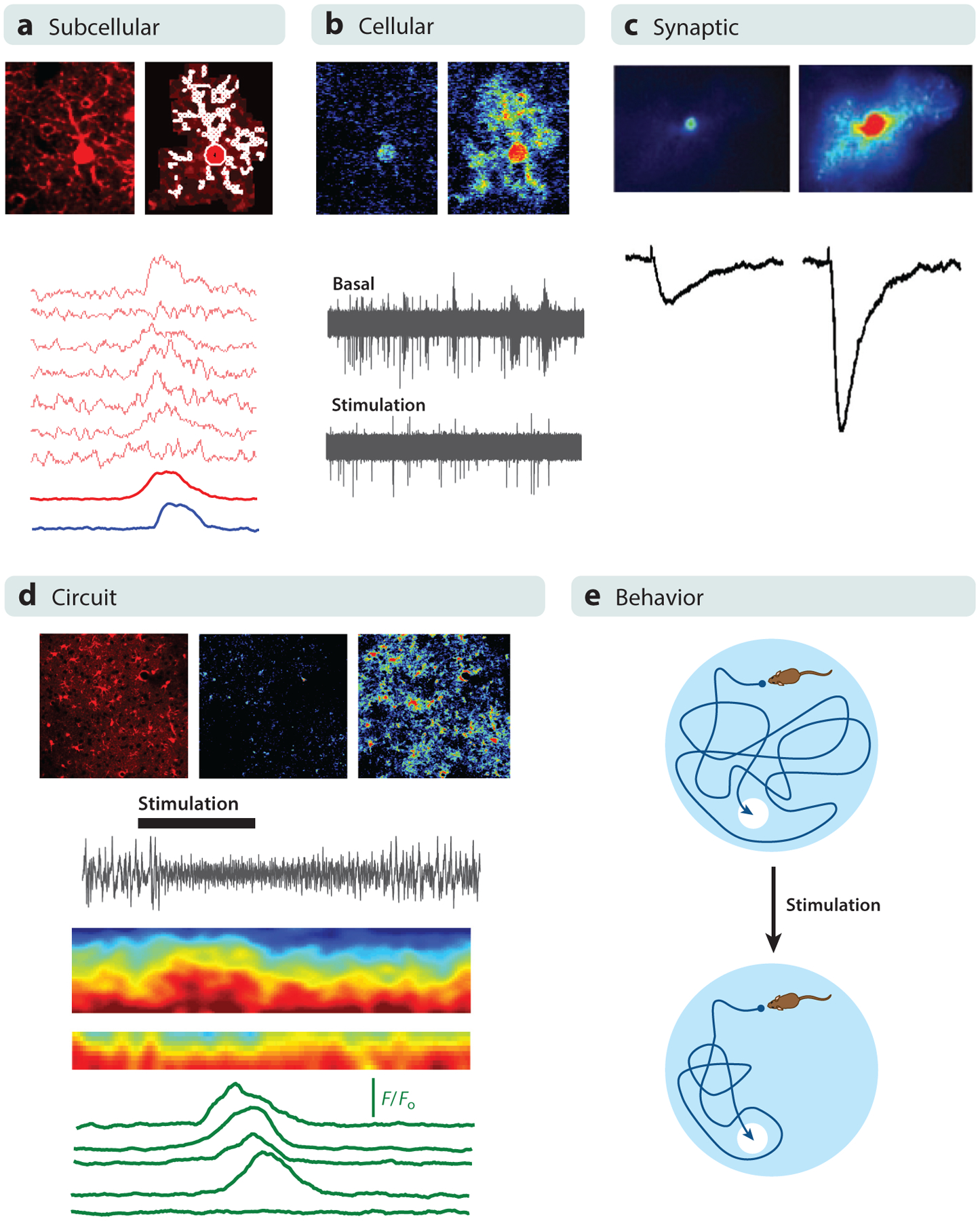
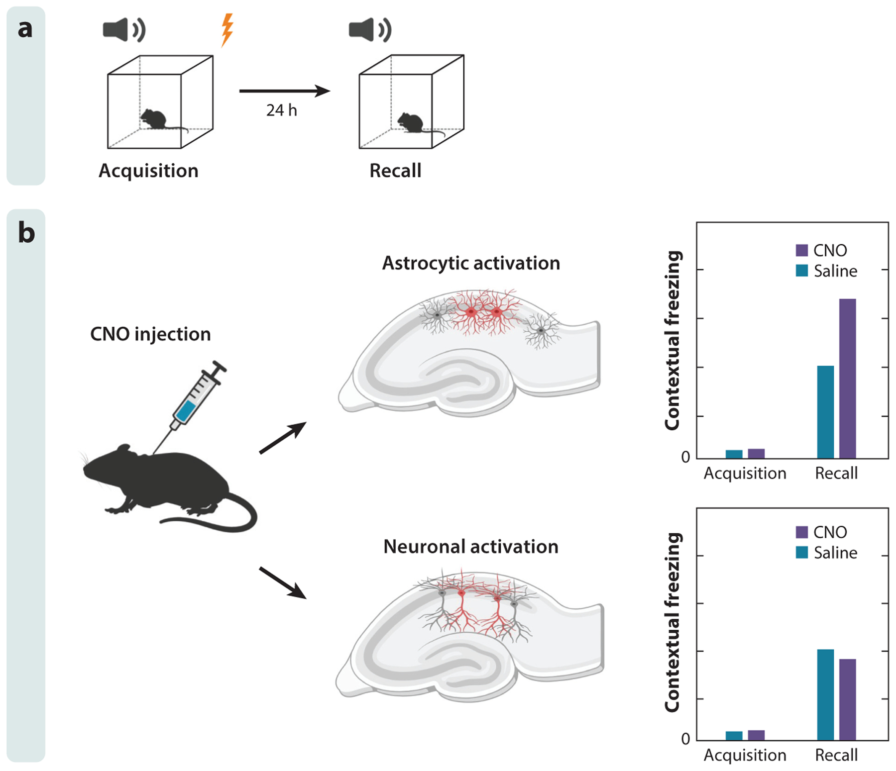
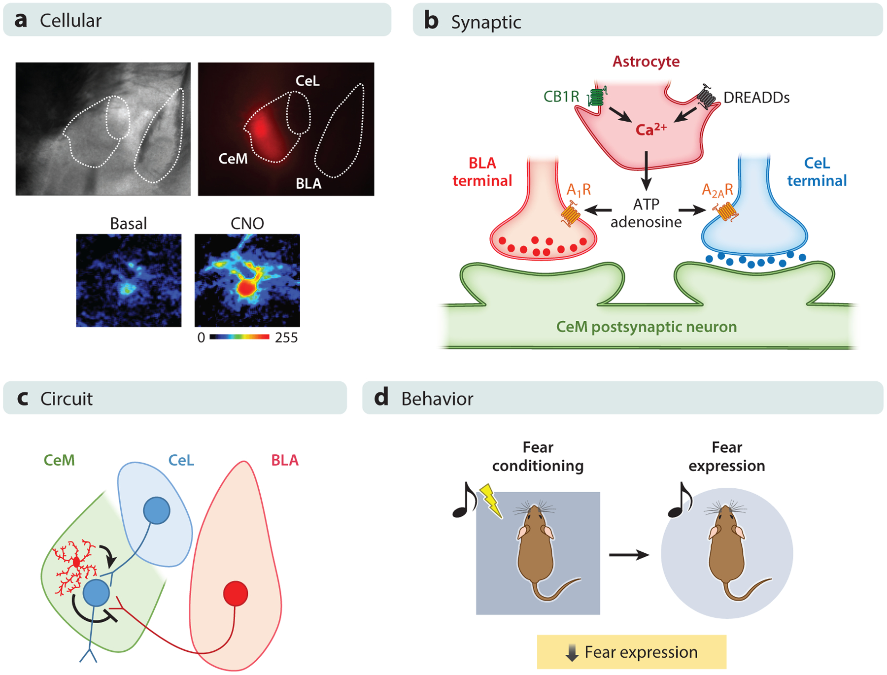
Animal behavior was classically considered to be determined exclusively by neuronal activity, whereas surrounding glial cells such as astrocytes played only supportive roles. However, astrocytes are as numerous as neurons in the mammalian brain, and current findings indicate a chemically based dialog between astrocytes and neurons. Activation of astrocytes by synaptically released neurotransmitters converges on regulating intracellular Ca2+ in astrocytes, which then can regulate the efficacy of near and distant tripartite synapses at diverse timescales through gliotransmitter release. Here, we discuss recent evidence on how diverse behaviors are impacted by this dialog. These recent findings support a paradigm shift in neuroscience, in which animal behavior does not result exclusively from neuronal activity but from the coordinated activity of both astrocytes and neurons. Decoding how astrocytes and neurons interact with each other in various brain circuits will be fundamental to fully understanding how behaviors originate and become dysregulated in disease.

Keywords: Ca2+ signaling; astrocytes; behavior; glia; gliotransmission; tripartite synapse.


Figures

Figure 1
Figure 1
Astrocyte activity and astrocyte-neuron interactions at different levels of analysis. (a) At the subcellular level, astrocytes display calcium elevations in microdomains. (Top) Images represent an SR101-labeled astrocyte (left) with marked microdomains (right); (bottom) traces represent calcium levels at microdomains in response to sensory stimulation. Panel a adapted with permission from Lines et al. (2020). (b) At the cellular level, astrocytes display generalized calcium elevations that impact neuronal electrical activity. (Top) Images represent pseudocolor calcium levels in a single astrocyte; (bottom) traces represent extracellular field recordings before and after astrocyte stimulation. Panel b adapted with permission from Lines et al. (2020). (c) At the synaptic level, calcium elevations in astrocytes regulate synaptic transmission properties. (Top) Images represent pseudocolor calcium levels in a single astrocyte; (bottom) traces represent synaptic currents in an adjacent single synapse before and after astrocyte stimulation. Panel c adapted with permission from Martin et al. (2015). (d) At the circuit level, astrocyte populations display generalized calcium elevations that influence neuronal network activity. (Top) Images represent SR101-labeled cortical astrocytes and pseudocolor calcium levels in an astrocyte population before and after sensory stimulation; (bottom) electrocorticogram trace, color-coded spectrogram, and traces represent astrocyte calcium levels before and after sensory stimulation. Panel d adapted with permission from Lines et al. (2020). (e) At the behavioral level, astrocyte-neuron interactions may influence multiple animal behaviors such as spatial learning of a hidden platform in a Morris water maze.
Figure 2
Figure 2
Astrocyte activation in the hippocampus is sufficient to induce memory enhancement. (a) A contextual fear conditioning paradigm is used to assess fear learning. Mice are exposed to electric shock and auditory cue during fear acquisition. Fear learning or recall is assessed by the freezing behavior displayed on exposure to auditory cue 24 h following fear acquisition. (b) Activation of CA1 astrocytes or neurons 30 min before fear acquisition has differential effects on fear learning. Chemogenetic activation of astrocytes in the mouse CA1 area of the hippocampus enhances fear learning, while chemogenetic activation of neuronal cells diminishes fear learning. Figure adapted with permission from Adamsky et al. (2018).
Figure 3
Figure 3
Astrocyte activity in the amygdala influences fear expression, which has been investigated at several levels in the central medial amygdala (CeM). (a) At the cellular level, expression of designer receptors exclusively activated by designer drugs (DREADDs) using clozapine-N-oxide (CNO) elicits calcium responses. (Top) Brightfield and fluorescence images show the localization of DREADDs in the CeM as reported by mCherry expression. (Bottom) Pseudocolor images of fluorescence intensities of a calcium indicator before and after local application of CNO are shown. (b) At the synaptic level, electrophysiological experiments show that the activation of astrocytes in the CeM, either by an endogenous stimulus or by a selective exogenous stimulus (DREADDs activation by CNO), is able to activate astrocytes and induce a regulation in central lateral amygdala (CeL)-CeM and basolateral amygdala (BLA)-CeM synapses. The astrocytic activity increases the synaptic probability of release in inhibitory CeL-CeM synapses through activation of A2A receptors and decreases the synaptic probability of release through activation of A1 receptors in excitatory BLA-CeM synapses. (c) At the circuit level, the combined and synergistic synaptic effects elicited upon astrocytic activation decrease the firing rate of CeM neurons. (d) At the behavioral level, the decrease of excitability of output CeM neurons results in decreases of behavioral fear expression as assessed using the cued fear-conditioning paradigm, in which an auditory stimulus is paired with an unconditioned stimulus. The fear expression is then tested by presenting only the conditioned stimulus in a novel surrounding context. Figure adapted with permission from Martin-Fernandez et al. (2017).

References

    1. Adamsky A, Goshen I. 2018. Astrocytes in memory function: pioneering findings and future directions. Neuroscience 370:14–26 - PubMed
    1. Adamsky A, Kol A, Kreisel T, Doron A, Ozeri-Engelhard N, et al. 2018. Astrocytic activation generates de novo neuronal potentiation and memory enhancement. Cell 174:59–71.e14 - PubMed
    1. Andersen JD, Pouzet B. 2004. Spatial memory deficits induced by perinatal treatment of rats with PCP and reversal effect of d-serine. Neuropsychopharmacology 29:1080–90 - PubMed
    1. Araque A, Carmignoto G, Haydon PG, Oliet SH, Robitaille R, Volterra A. 2014. Gliotransmitters travel in time and space. Neuron 81:728–39 - PMC - PubMed
    1. Arrigoni E, Chee MJS, Fuller PM. 2019. To eat or to sleep: That is a lateral hypothalamic question. Neuropharmacology 154:34–49 - PubMed

Publication types

LinkOut - more resources