. 2021 Jan 6;13(1):1440-1457.
doi: 10.18632/aging.202379. Epub 2021 Jan 6.

NLRC4 gene silencing-dependent blockade of NOD-like receptor pathway inhibits inflammation, reduces proliferation and increases apoptosis of dendritic cells in mice with septic shock

Affiliations

NLRC4 gene silencing-dependent blockade of NOD-like receptor pathway inhibits inflammation, reduces proliferation and increases apoptosis of dendritic cells in mice with septic shock

Shi-Sheng Wang et al. Aging (Albany NY). .

Abstract

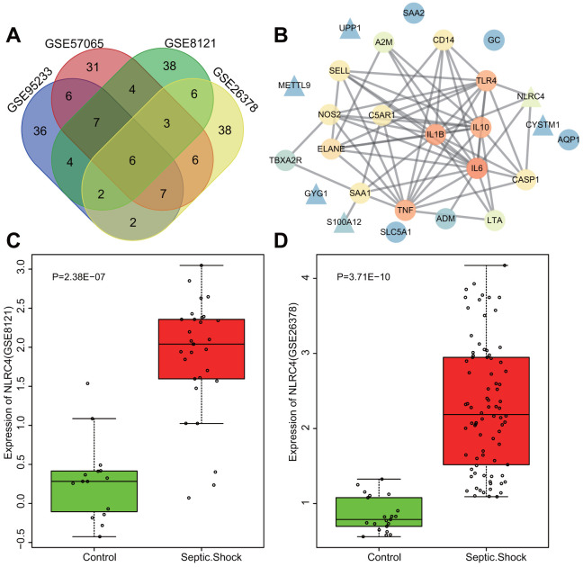
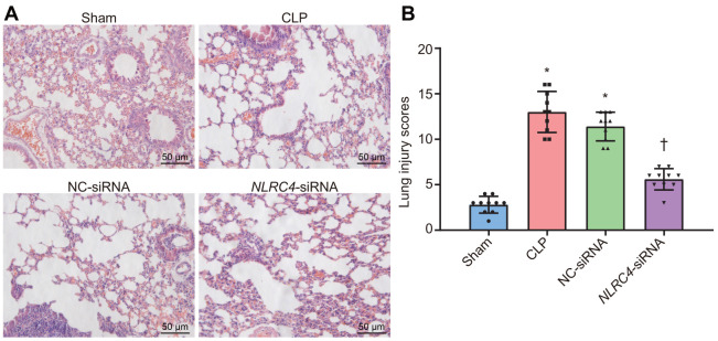
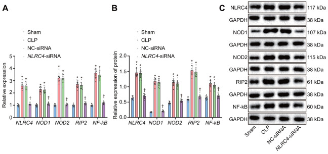
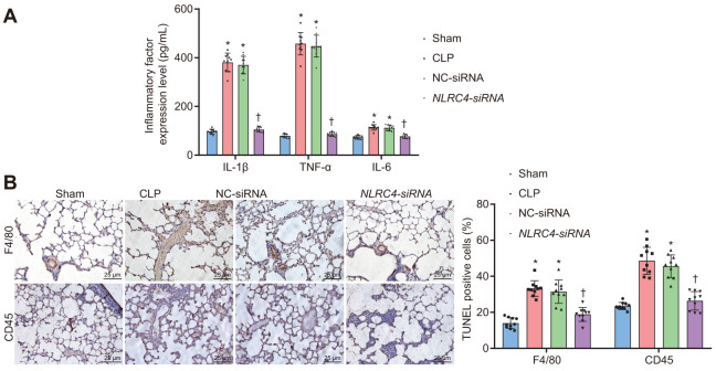
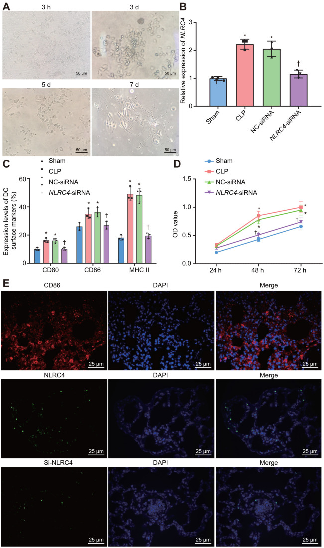
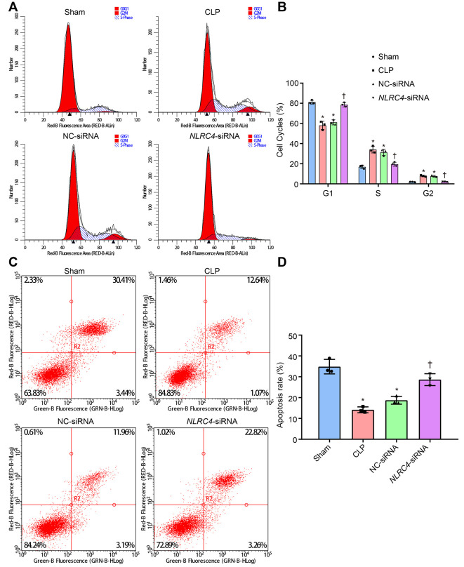
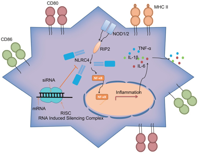
Septic shock is one of the most significant health concerns across the world, involving hypo-perfusion and defects in tissue energy. The current study investigates the role of NLR family CARD domain containing protein 4 (NLRC4) in septic shock-induced inflammatory reactions, lung tissue injuries, and dendritic cell (DC) apoptosis. Septic shock mice models were established by modified cecal ligation and puncture and injected with retroviral vector expressing siRNA-NLRC4. DCs were then isolated and transfected with siRNA-NLRC4. The degree of lung tissue injury, cell cycle distribution, cell apoptosis and cell viability of DCs were assessed. NLRC4 was found to be expressed at high levels in mice with septic shock. NLRC4 silencing inhibited the activation of the NOD-like receptor (NLR) pathway as evidenced by the decreased levels of NOD1, NOD2, RIP2, and NF-κB. In addition, NLRC4 silencing reduced the inflammatory reaction as attributed by reduced levels of IL-1β, TNF-α and IL-6. Suppressed NLRC4 levels inhibited cell viability and promoted cell apoptosis evidenced by inhibited induction of DC surface markers (CD80, CD86, and MHC II), along with alleviated lung tissue injury. In conclusion, NLRC4 silencing ameliorates lung injury and inflammation induced by septic shock by negatively regulating the NLR pathway.

Keywords: NLRC4; NOD-like receptor pathway; immune response; inflammatory reaction; septic shock.


Conflict of interest statement

CONFLICTS OF INTEREST: The authors declare that they have no conflicts of interest.

Figures

Figure 1
Figure 1
Highly expressed NLRC4 was found in septic shock. (A) 6 intersected genes detected among the top 70 DEGs from the microarray expression profiles of GSE95233, GSE57065, GSE8121 and GSE26378. (B) PPI network of the DEGs and septic shock-related genes (the triangle signifies the DEGs, the circle indicates the septic shock-related genes; the color of the genes shows the correlation degree with other genes, with orange coloration indicative of a high correlation degree and a blue coloration signifying a low correlation degree). (C, D) The expression of NLRC4 in septic microarray expression profiles GSE8121 and GSE26378.
Figure 2
Figure 2
HE staining results illustrate the amelioration of lung tissue injury induced by septic shock in mice after DCs transfected with NLRC4-siRNA. (A) The lung tissue injury detected by HE staining (scale bar = 50 μm). (B) The lung tissue injury scores. n = 10. * p < 0.05 vs. the sham group. † p < 0.05 vs. the CLP and NC-siRNA groups. Data comparison among multiple groups was analyzed by one-way analysis of variance (ANOVA), followed by Tukey’s post hoc test. The experiment was repeated 3 times independently.
Figure 3
Figure 3
Silencing of NLRC4 inactivates the NLR pathway in mouse lung tissues. (A) The mRNA expression of NLRC4, NOD1, NOD2, RIP2, and NF-κB in DCs determined by RT-qPCR. (B) The protein expression of NLRC4, NOD1, NOD2, RIP2, and NF-κB normalized to GAPDH in DCs measured by Western blot analysis. (C) Western blot bands of NLRC4, NOD1, NOD2, RIP2, and NF-κB in different transfection groups. N = 10. * p < 0.05 vs. the sham group. † p < 0.05 vs. the CLP and NC-siRNA groups. Data comparison among multiple groups was analyzed by one-way ANOVA, followed by Tukey’s post hoc test. The experiment was repeated 3 times independently.
Figure 4
Figure 4
NLRC4 downregulation alleviates inflammatory reaction and reduces inflammatory cell infiltration. (A) the levels of IL-1β, TNF-α and IL-6 in lung tissues of mice with septic shock detected by ELISA. (B) inflammatory cell infiltration evidenced by F4/80 and CD45 positive levels using TUNEL staining (scale bar = 25 μm). n = 10. * p < 0.05 vs. the sham group. † p < 0.05 vs. the CLP and NC-siRNA groups. Data comparison among multiple groups was analyzed by one-way ANOVA, followed by Tukey’s post hoc test. The experiment was repeated 3 times independently.
Figure 5
Figure 5
Improved morphological changes in DCs after being cultured for 3 h, 3 d, 5 d, and 7 d and suppressed NLRC4 reduces DC proliferation. (A) Morphological changes of DCs observed under an inverted microscope (scale bar = 50 μm). (B) The expression of NLRC4 in DCs determined by RT-qPCR. (C) The induction of DC surface markers, CD80, CD86, and MHC II, detected by flow cytometry. (D) The DC apoptosis as indicative of OD values detected by flow cytometry. (E) The immunofluorescence labeling of NLRC4 and DC surface marker CD86 (scale bar = 25 μm). The bone marrow-derived DCs from the same group of mice were adopted in the in vivo experiments. * p < 0.05 vs. the sham group. † p < 0.05 vs. the CLP and NC-siRNA groups. Data comparison among multiple groups was analyzed by one-way ANOVA, followed by Tukey’s post-hoc test. Data at different time points were compared by repeated measures ANOVA, followed by Bonferroni test. The experiment was repeated 3 times independently.
Figure 6
Figure 6
siRNA-mediated silencing of NLRC4 elevates DC cell apoptosis rate in mice with septic shock as evidenced by PI single staining and AnnexinV-FITC/PI double staining. (A, B), DC cycle distribution in the sham, CLP, NC-siRNA, and NLRC4-siRNA groups detected by PI single staining. (C, D) DC apoptosis in the sham, CLP, NC-siRNA, NLRC4-siRNA groups revealed by AnnexinV-FITC/PI double staining. The bone marrow-derived DCs from the same group of mice were adopted in the in vivo experiments. * p < 0.05 vs. the sham group. † p < 0.05 vs. the CLP and NC-siRNA groups. Data comparison among multiple groups was analyzed by one-way ANOVA, followed by Tukey’s post hoc test. The experiment was repeated 3 times independently.
Figure 7
Figure 7
NLRC4 gene regulates inflammatory reaction and immune response of DCs in septic shock by mediating the NLR pathway. NLRC4 is the receptor of the NLR pathway. Activated NF-κB pathway induces inflammatory reaction with secretion of inflammatory factors: IL-1β, TNF-α and IL-6 outside cells, as well as increases in CD80, CD86 and MHC II on the cytomembrane, thus activating DC immune response. Importantly, siRNA-mediated silencing of NLRC4 blocks the NLR signaling pathway to inhibit DC maturation and immune response, which alleviates the lung tissue injury of mice induced by septic shock.

Similar articles

Cited by

References

    1. Oriol I, Sabé N, Melilli E, Lladó L, González-Costello J, Soldevila L, Carratalà J. Factors influencing mortality in solid organ transplant recipients with bloodstream infection. Clin Microbiol Infect. 2015; 21:1104.e9–14. 10.1016/j.cmi.2015.07.021 - DOI - PubMed
    1. Vazquez-Grande G, Kumar A. Optimizing antimicrobial therapy of sepsis and septic shock: focus on antibiotic combination therapy. Semin Respir Crit Care Med. 2015; 36:154–66. 10.1055/s-0034-1398742 - DOI - PubMed
    1. Jessen KM, Lindboe SB, Petersen AL, Eugen-Olsen J, Benfield T. Common TNF-alpha, IL-1 beta, PAI-1, uPA, CD14 and TLR4 polymorphisms are not associated with disease severity or outcome from gram negative sepsis. BMC Infect Dis. 2007; 7:108. 10.1186/1471-2334-7-108 - DOI - PMC - PubMed
    1. Meyer NJ, Ferguson JF, Feng R, Wang F, Patel PN, Li M, Xue C, Qu L, Liu Y, Boyd JH, Russell JA, Christie JD, Walley KR, Reilly MP. A functional synonymous coding variant in the IL1RN gene is associated with survival in septic shock. Am J Respir Crit Care Med. 2014; 190:656–64. 10.1164/rccm.201403-0586OC - DOI - PMC - PubMed
    1. Holmes CL, Russell JA, Walley KR. Genetic polymorphisms in sepsis and septic shock: role in prognosis and potential for therapy. Chest. 2003; 124:1103–15. 10.1378/chest.124.3.1103 - DOI - PubMed