. 2021 Jan 4;13(1):53.
doi: 10.3390/pharmaceutics13010053.

Effect of Dexamethasone-Loaded PLGA Nanoparticles on Oral Mucositis Induced by 5-Fluorouracil

Affiliations

Effect of Dexamethasone-Loaded PLGA Nanoparticles on Oral Mucositis Induced by 5-Fluorouracil

Susana Barbosa Ribeiro et al. Pharmaceutics. .

Abstract

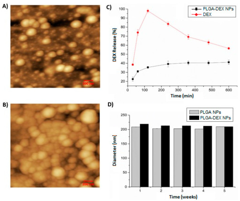
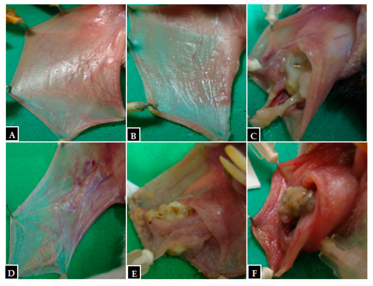
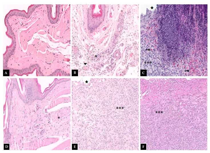
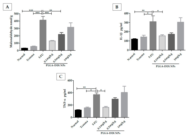
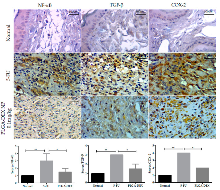
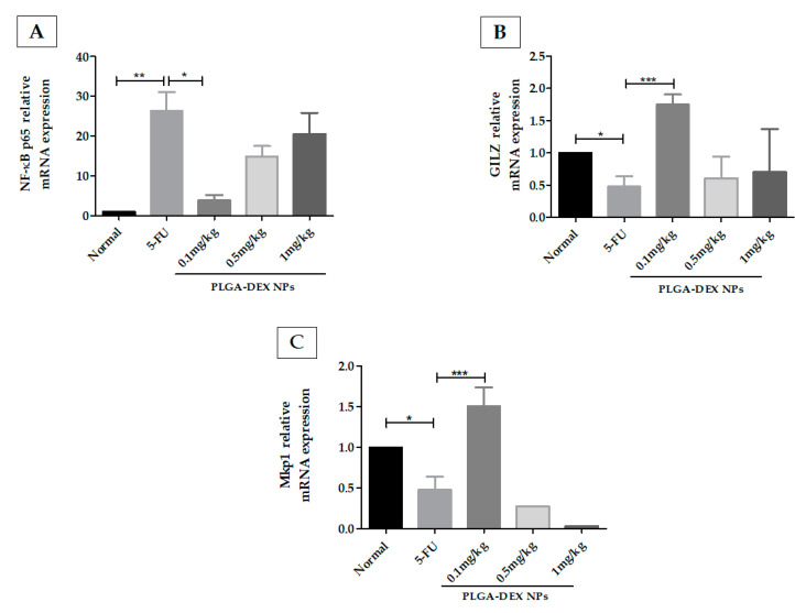
Oral mucositis (OM) is characterized by the presence of severe ulcers in the oral region that affects patients treated with chemotherapy. It occurs in almost all patients who receive radiotherapy of the head and neck, as well as patients who undergo hematopoietic cell transplantation. The pathophysiology of OM is complex, and there is no effective therapy. The aim of this study was to evaluate the effect of dexamethasone-loaded poly(d,l-Lactic-co-glycolic) nanoparticles (PLGA-DEX NPs) on an OM model induced in hamsters. The NPs were synthesized using the emulsification-solvent evaporation method and were characterized by the size, zeta potential, encapsulation efficiency, atomic force microscopy, physicochemical stability, and the in vitro release. The OM was induced by the administration of 5-FU on the first and second days and mechanical trauma on the 4th day of the experiment. PLGA-DEX NPs were administered to treat OM. The animals were euthanized on the 10th day. Macroscopic and histopathological analyses were performed, measurement of malonaldehyde (MDA) and ELISA was used to determine the levels of IL-1β and TNF-α. Immunoexpressions of NF-κB, COX-2, and TGF-β were determined by immunohistochemistry, and qRT-PCR was used to quantify the gene expression of the GILZ, MKP1, and NF-κB p65. The PLGA-DEX NPs (0.1 mg/kg) significantly reduced macroscopic and histopathological scores, decreased MDA, TNF-α and IL-1β levels, immunostaining for NF-κB, COX-2, TGF-β, and suppressed NF-κB p65 mRNA expression, but increased GILZ and MKP1 expression.

Keywords: 5-fluorouracil; PLGA; nanoparticles; oral mucositis.


Conflict of interest statement

The authors declare no conflict of interest. The funders had no role in the design of the study; in the collection, analyses, or interpretation of data; in the writing of the manuscript, or in the decision to publish the results.

Figures

Figure 1
Figure 1
Atomic force microscopy (AFM)—2D images—200 nm scale. (A) The nanoparticle of poly(d, Lactic-co-glycolic) nanoparticles (PLGA NP). (B) dexamethasone-loaded poly (d, Lactic-co-glycolic) nanoparticles (PLGA-DEX NP). (C) In vitro release profile of dexamethasone free in solution (DEX) and PLGA-DEX NP. (D) Physical stability of PLGA NP and PLGA-DEX NP for 5 weeks.
Figure 2
Figure 2
Macroscopic images from hamster oral mucosa with oral mucositis (OM) induced by 5-fluorouracil (5-FU) and mechanical trauma (MT), treated with PLGA-DEX NPs. (A) Normal group: animals not subjected to induction of OM received daily administrations of purified water i.p. (B) Trauma group: animals subjected to MT only, without OM inductions, received purified water i.p. daily. (C) Group 5-FU: animals with OM induced by 5-FU and MT received purified water i.p. daily. PLGA-DEX NP groups (D) 0.1 mg/kg, (E) 0.5 mg/kg, (F) 1 mg/kg: animals with OM treated with daily administrations of PLGA-DEX NPs i.p. at the dose specified for each of the three groups.
Figure 3
Figure 3
Histopathological examination of the hamster oral mucosa. OM was induced by 5-FU and MT, treated with PLGA-DEX NPs. (A) Normal group: animals without histopathological changes in oral mucosa. (B) Trauma group: animals subjected to the induction of excoriations in the oral mucosa, without OM; there are blood vessels (triangle) and a small region of inflammatory infiltrate (an asterisk) dispersed in the conjunctive. (C) Group 5-FU: animals with untreated OM, characterized by the presence of ulcers (star), intense inflammatory infiltrate (three asterisks), and hemorrhagic foci (arrow) dispersed throughout the region. PLGA-DEX groups (D) 0.1 mg/kg, (E) 0.5 mg/kg, (F) 1 mg/kg: animals with OM treated with DEX-loaded PLGA NPs. The groups treated with PLGA-DEX NPs 0.5 and 1 mg/kg presented intense inflammatory infiltrate (three asterisk) and ulcers (star). The group treated with PLGA-DEX NPs 0.1 mg/kg presented a reduction in inflammation, marked by a decrease in inflammatory infiltrate (an asterisk).
Figure 4
Figure 4
Malonaldehyde (MDA) and cytokine quantifications in the hamster oral mucosa to evaluate the therapeutic outcome of PLGA-DEX NPs in OM induced by 5-FU. (A) MDA. (B) IL-1β. (C) TNF-α. Normal group: animals with healthy oral mucosa. Trauma group: animals subjected only to excoriations in the oral mucosa, but they do not present OM. Group 5-FU: animals with untreated OM only received purified water, i.p. The PLGA-DEX NPs groups (0.1; 0.5 or 1 mg/kg) received 5-FU and were subjected to MT with consequent OM induction, which was treated with PLGA-DEX NP i.p. in the dose corresponding to the group to which they belonged. The results are presented as the mean ± standard error (n = 5). * p < 0.05, ** p < 0.01, *** p < 0.001 (ANOVA with Tukey’s posttest).
Figure 5
Figure 5
Immunoexpression photomicrographs of NF-κB, TGF-β, and COX-2 in the normal, 5-FU, and PLGA-DEX 0.1 mg/kg groups. The immunostaining of these proteins was greater in the 5-FU group, while the normal and PLGA-DEX NPs groups exhibited low expression, bars indicating 100 µm. Expression scores for NF-κB, TGF-β, and COX-2 with a 95% confidence interval. * p < 0.05; ** p < 0.01 (Kruskal–Wallis test, followed by Dunn’s test for post hoc comparisons).
Figure 6
Figure 6
(A) q-RT PCR for NF-kB p65, (B) glucocorticoid-induced leucine zipper (GILZ), (C) MAPK phosphatase 1 (MKP1). 5-FU increased the expression of the NF-κB p65 genes and decreased the gene expression of GILZ and MKP1, compared to the normal group. The animals in the PLGA-DEX group 0.1 mg/kg showed increased GILZ and MKP1 gene expression as well as reduced expression of the NF-κB p65 gene, compared to the 5-FU group (n = 5; * p < 0.05, ** p < 0.01, *** p < 0.001) (ANOVA followed by Tukey’s posttest).

References

    1. Sonis S. A biological approach to mucositis. J. Support. Oncol. 2004;2:21–36. - PubMed
    1. Barasch A., Peterson D.E. Risk factors for ulcerative oral mucositis in cancer patients: Unanswered questions. Oral Oncol. 2003;39:91–100. doi: 10.1016/S1368-8375(02)00033-7. - DOI - PubMed
    1. Elting L.S., Keefe D.M., Sonis S.T., Garden A.S., Spijkervet F.K.L., Barasch A., Tishler R.B., Canty T.P., Kudrimoti M.K., Vera-Llonch M., et al. Patient-reported measurements of oral mucositis in head and neck cancer patients treated with radiotherapy with or without chemotherapy: Demonstration of increased frequency, severity, resistance to palliation, and impact on quality of life. Cancer. 2008;113:2704–2713. doi: 10.1002/cncr.23898. - DOI - PubMed
    1. Rastogi M., Dwivedi R., Kazi R. Oral mucositis in head and neck cancer. Eur. J. Cancer Care. 2011;20:144. doi: 10.1111/j.1365-2354.2011.01243.x. - DOI - PubMed
    1. Ribeiro S.B., De Araújo A.A., De Araújo Júnior R.F., De Castro Brito G.A., Leitão R.C., Barbosa M.M., Garcia V.B., Medeiros A.C., De Medeiros C.A.C.X. Protective effect of dexamethasone on 5-FU-induced oral mucositis in hamsters. PLoS ONE. 2017;12:e0186511. doi: 10.1371/journal.pone.0186511. - DOI - PMC - PubMed

LinkOut - more resources