Review
. 2021 Jan 4;26(1):217.
doi: 10.3390/molecules26010217.

Recent Advances in Organelle-Targeted Fluorescent Probes

Affiliations
Review

Recent Advances in Organelle-Targeted Fluorescent Probes

Na-Eun Choi et al. Molecules. .

Abstract

Recent advances in fluorescence imaging techniques and super-resolution microscopy have extended the applications of fluorescent probes in studying various cellular processes at the molecular level. Specifically, organelle-targeted probes have been commonly used to detect cellular metabolites and transient chemical messengers with high precision and have become invaluable tools to study biochemical pathways. Moreover, several recent studies reported various labeling strategies and novel chemical scaffolds to enhance target specificity and responsiveness. In this review, we will survey the most recent reports of organelle-targeted fluorescent probes and assess their general strategies and structural features on the basis of their target organelles. We will discuss the advantages of the currently used probes and the potential challenges in their application as well as future directions.

Keywords: chemical probes; fluorescence imaging; organelle-targeting.


Conflict of interest statement

The authors declare no conflict of interest.

Figures

Figure 1
Figure 1
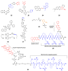
(a) Molecular design of 6 and proposed sensing mechanism; (b) Schematic illustration of in situ response of 6 controlled by UV irradiation. Reprinted with permission from ref. [13]. Copyright 2020 American Chemical Society.
Figure 2
Figure 2
Lysosome-targeted fluorescent probes (blue: targeting moiety; red: responsive moiety; orange: photocaging group).
Figure 3
Figure 3
(a) Structures of nucleus-targeted probes (blue: targeting moiety; red: responsive moiety; orange: photocaging group); (b) Structure of 13 (SiR-TTet59B) and a schematic representation of fluorogenic recognition of the telomere sequence. Reprinted with permission from ref. [30]. Copyright 2020 American Chemical Society; (c) Schematic illustration of 14 targeting the nucleus of integrin ανβ3 and CD13-overexpressed cancer cells. Reprinted with permission from ref. [31]—Published by The Royal Society of Chemistry.
Figure 4
Figure 4
(a) A: Amphiphilic NIR-AZA probe (18); B: Simplified schematic showing representation of membrane DIE activation of aggregated 18. Reprinted with permission from ref. [41], copyright Elsevier 2018; (b) Structures of membrane-targeted probes (blue: targeting moiety; red: responsive moiety).
Figure 5
Figure 5
Morphological changes of the mitochondrial inner membrane captured by S stimulated emission depletion (STED) microscopy using 26. (A): Deconvoluted STED images showing changes in the mitochondrial morphology; (B): Comparison of the number of cristae per micrometer of mitochondrial length before and after incubation for 3 and 12 h under starvation conditions; (C): STED image of cristae in HeLa cells, pretreated with 10 µM mitochondrial DNA replication inhibitor (ddC) for 5 days followed by staining with MitoPB Yellow. Reprinted with permission from ref. [62].
Figure 6
Figure 6
(A) Aptamer/DQAsome-based mitochondrion-targeted probe (28) and schematic of DNA probe/DQAsome complex formulation. (B) Schematic depiction of targeted delivery of DNA probe/DQAsomes to mitochondria. Reprinted with permission from ref. [65]—Published by The Royal Society of Chemistry.
Figure 7
Figure 7
Mitochondrion-targeted probes (blue: targeting moiety; red: responsive moiety; orange: photocaging group).
Figure 8
Figure 8
Endoplasmic reticulum (ER)- and Golgi-targeted probes (blue: targeting moiety; red: responsive moiety).
Figure 9
Figure 9
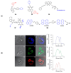
(a) Structures of probes targeting melanosomes (3435), peroxisomes (36), and lipid droplets (LD) (3738) (blue: targeting moiety; red: responsive moiety); (b) Fluorescence imaging of brown adipocytes stained with Lipi-probes (38)—Lipi-Blue (A–C), Lipi-Green (D–F), Lipi-Red (G–I). Images of differential interference contrast. (B, E, and H) Images of fluorescence. (C, F, and I) Images of overlay. The graphs on the right are results of the line-scan analysis: B, Lipi-Blue; E, Lipi-Green; or H, Lipi-Red. (J, K, and L) Fluorescence intensity of the range indicated by the arrows shown in parts B, E, and H, respectively. Scale bar is 10 μm. Reprinted with permission from ref. [97]. Copyright 2019 American Chemical Society.

References

    1. Perera R.M., Zoncu R. The Lysosome as a Regulatory Hub. Annu. Rev. Cell Dev. Biol. 2016;32:223–253. doi: 10.1146/annurev-cellbio-111315-125125. - DOI - PMC - PubMed
    1. Xu H., Ren D. Lysosomal physiology. Annu. Rev. Physiol. 2015;77:57–80. doi: 10.1146/annurev-physiol-021014-071649. - DOI - PMC - PubMed
    1. Bonam S.R., Wang F., Muller S. Lysosomes as a therapeutic target. Nat. Rev. Drug Discov. 2019;18:923–948. doi: 10.1038/s41573-019-0036-1. - DOI - PMC - PubMed
    1. Deus C.M., Yambire K.F., Oliveira P.J., Raimundo N. Mitochondria-Lysosome Crosstalk: From Physiology to Neurodegeneration. Trends Mol. Med. 2020;26:71–88. doi: 10.1016/j.molmed.2019.10.009. - DOI - PubMed
    1. Hastings K.T., Cresswell P. Disulfide reduction in the endocytic pathway: Immunological functions of gamma-interferon-inducible lysosomal thiol reductase. Antioxid. Redox Signal. 2011;15:657–668. doi: 10.1089/ars.2010.3684. - DOI - PMC - PubMed

Substances

LinkOut - more resources