. 2021 Jan 4;22(1):434.
doi: 10.3390/ijms22010434.

Inhibition of RANKL-Induced Osteoclastogenesis by Novel Mutant RANKL

Affiliations

Inhibition of RANKL-Induced Osteoclastogenesis by Novel Mutant RANKL

Yuria Jang et al. Int J Mol Sci. .

Abstract

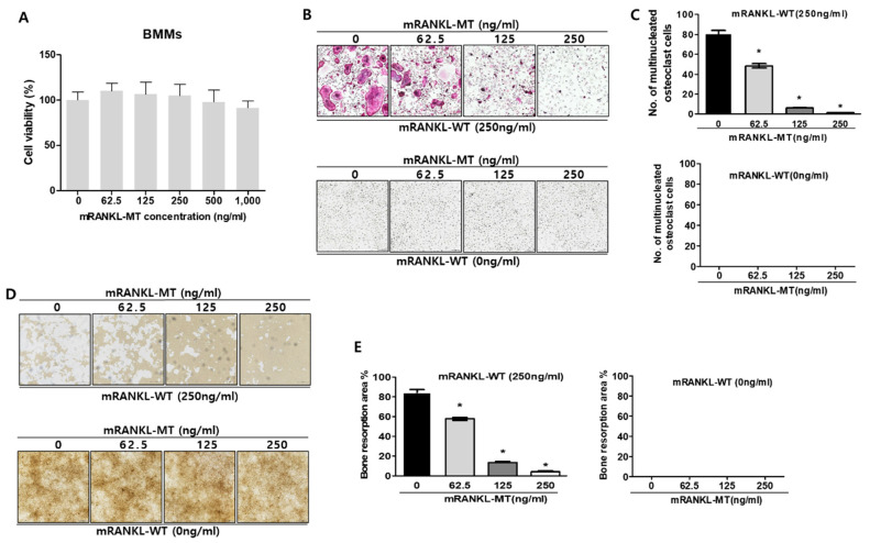
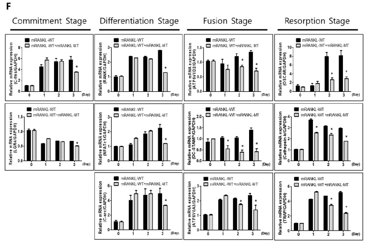
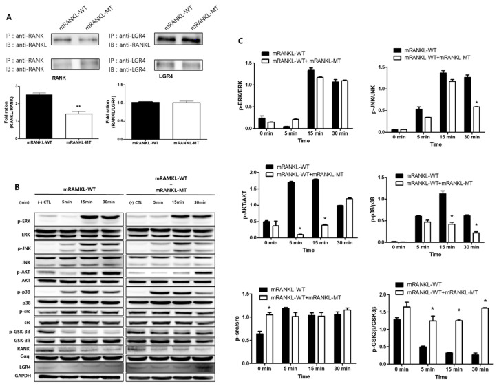
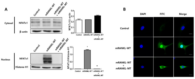
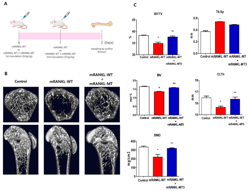
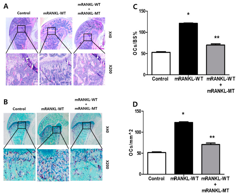
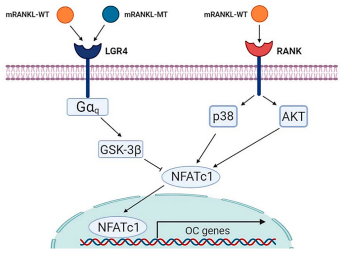
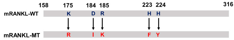
Background: Recently, it was reported that leucine-rich repeat-containing G-protein-coupled receptor 4 (LGR4, also called GPR48) is another receptor for RANKL and was shown to compete with RANK to bind RANKL and suppress canonical RANK signaling during osteoclast differentiation. The critical role of the protein triad RANK-RANKL in osteoclastogenesis has made their binding an important target for the development of drugs against osteoporosis. In this study, point-mutations were introduced in the RANKL protein based on the crystal structure of the RANKL complex and its counterpart receptor RANK, and we investigated whether LGR4 signaling in the absence of the RANK signal could lead to the inhibition of osteoclastogenesis.; Methods: The effects of point-mutated RANKL (mRANKL-MT) on osteoclastogenesis were assessed by tartrate-resistant acid phosphatase (TRAP), resorption pit formation, quantitative real-time polymerase chain reaction (qPCR), western blot, NFATc1 nuclear translocation, micro-CT and histomorphological assay in wild type RANKL (mRANKL-WT)-induced in vitro and in vivo experimental mice model.

Results: As a proof of concept, treatment with the mutant RANKL led to the stimulation of GSK-3β phosphorylation, as well as the inhibition of NFATc1 translocation, mRNA expression of TRAP and OSCAR, TRAP activity, and bone resorption, in RANKL-induced mouse models; and Conclusions: The results of our study demonstrate that the mutant RANKL can be used as a therapeutic agent for osteoporosis by inhibiting RANKL-induced osteoclastogenesis via comparative inhibition of RANKL. Moreover, the mutant RANKL was found to lack the toxic side effects of most osteoporosis treatments.

Keywords: leucine-rich repeat-containing G-protein-coupled receptor 4; osteoclast; osteoporosis; receptor activator of nuclear factor kappa-Β ligand.


Conflict of interest statement

The authors declare no conflict of interest.

Figures

Figure 1
Figure 1
Effect of mutant RANKL (mRANKL-MT) on osteoclast differentiation in vitro (A). Cell viability was determined by 3-(4,5-dimethylthiazol-2-yl)-2,5-diphenyltetrazolium bromide (MTT) assay. BMMs cells were exposed to various doses (0, 62.5, 125, 250, 500 and 1000 ng/mL) of mutant RANKL. The results are presented as the mean ± SD of three independent experiments, and there shows no significant differences (p < 0.05) compared to the 0 ng/mL mutant RANKL-treated group. (B) A typical image of BMMs stained for TRAP (red) after treatment with various doses of mRANKL-MT (0, 62.5, 125, 250 ng) in the presence or absence of mRANKL-WT (250 ng/mL). (C). Numbers of multinucleated TRAP cells (BMMs) (red) (≥3 nuclei) in these cultures (n = 4); (D). BMMs were incubated in hydroxyapatite-coated plates with various doses of mRANKL-MT (0, 62.5, 125, 250 ng) in the presence or absence of mRANKL-WT (250 ng/mL). The cells attached to the plate were removed and imaged using a light microscope (E). The absorption area was quantified using the Image J software. * p < 0.01. (F) mRANKL-MT inhibits RANKL-induced osteoclast gene expression. BMMs were exposed to mRANKL-WT (2 µg/mL) or mRANKL-WT (2 µg/mL) + mRANKL-MT (2 µg/mL) for 3 days. Gene expression was determined by real-time PCR and normalized to the expression of GAPDH. The data come from three separate experiments and are expressed as the mean ± standard deviation (SD). * p < 0.01.
Figure 1
Figure 1
Effect of mutant RANKL (mRANKL-MT) on osteoclast differentiation in vitro (A). Cell viability was determined by 3-(4,5-dimethylthiazol-2-yl)-2,5-diphenyltetrazolium bromide (MTT) assay. BMMs cells were exposed to various doses (0, 62.5, 125, 250, 500 and 1000 ng/mL) of mutant RANKL. The results are presented as the mean ± SD of three independent experiments, and there shows no significant differences (p < 0.05) compared to the 0 ng/mL mutant RANKL-treated group. (B) A typical image of BMMs stained for TRAP (red) after treatment with various doses of mRANKL-MT (0, 62.5, 125, 250 ng) in the presence or absence of mRANKL-WT (250 ng/mL). (C). Numbers of multinucleated TRAP cells (BMMs) (red) (≥3 nuclei) in these cultures (n = 4); (D). BMMs were incubated in hydroxyapatite-coated plates with various doses of mRANKL-MT (0, 62.5, 125, 250 ng) in the presence or absence of mRANKL-WT (250 ng/mL). The cells attached to the plate were removed and imaged using a light microscope (E). The absorption area was quantified using the Image J software. * p < 0.01. (F) mRANKL-MT inhibits RANKL-induced osteoclast gene expression. BMMs were exposed to mRANKL-WT (2 µg/mL) or mRANKL-WT (2 µg/mL) + mRANKL-MT (2 µg/mL) for 3 days. Gene expression was determined by real-time PCR and normalized to the expression of GAPDH. The data come from three separate experiments and are expressed as the mean ± standard deviation (SD). * p < 0.01.
Figure 2
Figure 2
Comparative inhibition of osteoclastogenesis by RANKL variant. (A) Co-immunoprecipitation for RANK or LGR4 binding RANKL variant in BMMs. Each blot was obtained under the same experimental conditions. The densitometric analysis is represented as the mean ratio ± standard deviation (SD) of three separate experiments. * p < 0.01, control group vs. mRANKL-WT group; ** p < 0.01 mRANKL-WT group vs. mRANKL-WT + mRANKL-MT group. (B) Western blot analysis of the RANK and LGR4 signaling cascades. GAPDH was used as a loading control. (C) A densitometric analysis of protein phosphorylation in ERK, JNK, AKT, P38, src and GSK-3β. Results are representative of three separate experiments with comparable results. BMMs were exposed to mRANKL-WT (2 µg/mL) or mRANKL-WT (2 µg/mL) + mRANKL-MT (2 µg/mL) for various time intervals.
Figure 3
Figure 3
The inhibitory effect of mRANKL-MT on NFATc1 translocation in BMMs. (A) NFATc1 nuclear translocation was analyzed by western blot in the cytoplasmic and nuclear fractions. Histone-H1 and β-actin were used as loading controls for the nuclear and cytoplasmic fractions, respectively. The densitometric analysis of NFATc1 in the cytoplasmic and nuclear fractions is represented as the mean ratio ± standard deviation (SD) of three separate experiments. * p < 0.01, control group vs. mRANKL-WT group; ** p < 0.01 mRANKL-WT group vs. mRANKL-WT + mRANKL-MT group. (B) NFATc1 nuclear translocation under confocal microscopy. Immunofluorescence images were acquired by staining for NFATc1 (green) and the nucleus (blue). Magnifications are 200Χ. Size bar is 20 μm.
Figure 4
Figure 4
3D micro-computed tomography (micro-CT) images of mouse femurs (A). Time schedule for immunization and sampling in mRANKL-WT or mRANKL-WT+mRANKL-MT-treated mouse. (B) Representative X-ray and micro-CT images of the distal femurs of intact mice (control), RANKL-induced osteoporosis mice (mRANKL-WT), and mRANKL-WT-induced osteoporosis mice treated with mRANKL-MT (C). Bone volume/total volume (BV/TV), trabecular thickness (Tb.Th), trabecular spacing (Tb/sp), Bone Volume (BV), Cortical thickness (Ct/Th), Bone Mineral Density (BMD). * p < 0.01, control group vs. mRANKL-WT group; ** p < 0.01 mRANKL-WT group vs. mRANKL-WT + mRANKL-MT group.
Figure 5
Figure 5
mRANKL-MT decreases the formation of osteoclast in mice (A) Histomorphometric analysis images. Magnifications are 20×. Size bar is 500 μm. (B) TRAP staining images of the femurs. Magnifications are 100×. Size bar is 50 μm. (C,D) Parameters of the femur osteoclasts. Oc.S/BS, osteoclast surface per bone surface; Oc.N/BS, osteoclast number per bone surface. Values were expressed as means ± standard deviation (SD). * p < 0.01 control vs. mRANKL-WT, ** p < 0.01 mRANKL-WT vs. mRANKL-WT + mRANKL-MT.
Figure 6
Figure 6
Schematic diagram of mRANKL-MT in inhibitory effect against mRANKL-WT/RANK signaling during osteoclastogenesis.
Figure 7
Figure 7
Protein sequence of mutant RANKL (mtRANKL), including the K180R, D189I, R190K, H223F, and H224Y transformants.

References

    1. Tanaka Y., Nakayamada S., Okada Y. Osteoblasts and osteoclasts in bone remodeling and inflammation. Curr. Drug. Targets Inflamm. Allergy. 2005;4:325–328. doi: 10.2174/1568010054022015. - DOI - PubMed
    1. Landesberg R., Woo V., Cremers S., Cozin M., Marolt D., Vunjak-Novakovic G., Kousteni S., Raghavan S. Potential pathophysiological mechanisms in osteonecrosis of the jaw. Ann. N. Y. Acad. Sci. 2011;1218:62. doi: 10.1111/j.1749-6632.2010.05835.x. - DOI - PMC - PubMed
    1. Feng X., McDonald J.M. Disorders of bone remodeling. Annu. Rev. Pathol. 2011;6:121–145. doi: 10.1146/annurev-pathol-011110-130203. - DOI - PMC - PubMed
    1. Bi H., Chen X., Gao S., Yu X., Xiao J., Zhang B., Liu X., Dai M. Key triggers of osteoclast-related diseases and available strategies for targeted therapies: A review. Front. Med. 2017;4:234. doi: 10.3389/fmed.2017.00234. - DOI - PMC - PubMed
    1. Hofbauer L., Kuhne C., Viereck V. The OPG/RANKL/RANK system in metabolic bone diseases. J. Musculoskelet. Neuronal Interact. 2004;4:268. - PubMed

LinkOut - more resources