Review
. 2021 Jan 4;22(1):433.
doi: 10.3390/ijms22010433.

Role of Peroxisome Proliferator-Activated Receptors (PPARs) in Trophoblast Functions

Affiliations
Review

Role of Peroxisome Proliferator-Activated Receptors (PPARs) in Trophoblast Functions

Lin Peng et al. Int J Mol Sci. .

Abstract

Peroxisome proliferator-activated receptors (PPARα, PPARβ/δ, and PPARγ) belong to the transcription factor family, and they are highly expressed in all types of trophoblast during pregnancy. The present review discusses currently published papers that are related to the regulation of PPARs via lipid metabolism, glucose metabolism, and amino acid metabolism to affect trophoblast physiological conditions, including differentiation, maturation, secretion, fusion, proliferation, migration, and invasion. Recent pieces of evidence have proven that the dysfunctions of PPARs in trophoblast lead to several related pregnancy diseases such as recurrent miscarriage, preeclampsia, intrauterine growth restriction, and gestational diabetes mellitus. Moreover, the underlying mechanisms of PPARs in the control of these processes have been discussed as well. Finally, this review's purposes are to provide more knowledge about the role of PPARs in normal and disturbed pregnancy with trophoblast, so as to find PPAR ligands as a potential therapeutic target in the treatment and prevention of adverse pregnancy outcomes.

Keywords: cytotrophoblast; extravillous trophoblast; functions; peroxisome proliferator-activated receptors (PPARs).


Conflict of interest statement

The authors declare no conflict of interest.

Figures

Figure 1
Figure 1
Illustrative representation of localization and mechanism of action of PPARs. PPARs belong to the nuclear receptor superfamily and consist of PPARα, PPARβ/δ, and PPARγ. Endogenous PPAR ligands can be transferred from the cytosol or generated in the nuclear membrane. PPARs bind to the specific DNA sequence AACT AGGNCA A AGGTCA, PPAR responsive element (PPRE), and their activation requires heterodimerization with another nuclear receptor, RXR. PPARs modulate numerous target genes by co-activator or co-inhibitor activity. PPARs are mainly expressed in the nucleus, and could shuttle from nuclear to cytoplasm. PPARγ transport from nuclear to cytoplasmic occurs via the extracellular signal-regulated kinase (ERK) cascade, a component of the mitogen-activated protein kinase (MAPK)/MEK 1/2. PPARs, peroxisome proliferator-activated receptors; RXR, 9-cis retinoic acid receptor.
Figure 2
Figure 2
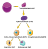
The path of human trophoblast differentiation. Trophoblast stem cells are derived from the trophectoderm. The cytotrophoblasts differentiate in two ways: invasive extravillous trophoblast (EVT) and syncytiotrophoblast (STB). EVT can be divided into two subtypes, one of which invades deeply into the uterine wall as groups of cells, termed interstitial EVT, and the other invades the maternal decidual arterioles as endovascular EVT.
Figure 3
Figure 3
PPARγ involved in trophoblast differentiation. The activation of PPARγ increased the expression of chorionic gonadotropin beta-subunit (hCGβ) and chorion-specific transcription factor (GCM-1), which promoted cytotrophoblast (CTB) differentiation into the STB. miR-1246 promoted STB differentiation through inhibiting the WNT/β-catenin signaling pathway and increasing the expression of the crucial transcription factor PPARγ. The loss of the ERK/MAPK cascade caused the reduced expression of PPARγ, affected the expression of GCM1, and inhibited the process of CTB differentiation into the STB.
Figure 4
Figure 4
PPARs linked hypoxia with trophoblast differentiation. The hypoxia inhibited the expression of PPARγ with the inactivation of both hypoxia inducible factor (HIF) and histone deacetylases (HDACs), which hampered the trophoblast differentiation. Hypoxia decreased the expression of TBP-2 and breast cancer resistance protein (BCRP) induced by suppressing PPARα and γ, which affected trophoblast differentiation. DHA increased the secretion of VEGF and the PPARβ ligand (GW501516) stimulated ANGPTL4 expression, which influenced the trophoblast differentiation.

Similar articles

Cited by

References

    1. Brunmeir R., Xu F. Functional Regulation of PPARs through Post-Translational Modifications. Int. J. Mol. Sci. 2018;19:1738. doi: 10.3390/ijms19061738. - DOI - PMC - PubMed
    1. Bensinger S.J., Tontonoz P. Integration of metabolism and inflammation by lipid-activated nuclear receptors. Nature. 2008;454:470–477. doi: 10.1038/nature07202. - DOI - PubMed
    1. Tsukahara T., Matsuda Y., Haniu H. Lysophospholipid-Related Diseases and PPARγ Signaling Pathway. Int. J. Mol. Sci. 2017;18:2730. doi: 10.3390/ijms18122730. - DOI - PMC - PubMed
    1. Bogacka I., Kurzynska A., Bogacki M., Chojnowska K. Peroxisome proliferator-activated receptors in the regulation of female reproductive functions. Folia Histochem. Cytobiol. 2015;53:189–200. doi: 10.5603/fhc.a2015.0023. - DOI - PubMed
    1. Holdsworth-Carson S.J., Lim R., Mitton A., Whitehead C., Rice G.E., Permezel M., Lappas M. Peroxisome proliferator-activated receptors are altered in pathologies of the human placenta: Gestational diabetes mellitus, intrauterine growth restriction and preeclampsia. Placenta. 2010;31:222–229. doi: 10.1016/j.placenta.2009.12.009. - DOI - PubMed

Substances

LinkOut - more resources