. 2021 Feb 2;93(4):2003-2009.
doi: 10.1021/acs.analchem.0c03173. Epub 2021 Jan 6.

N- and O-Glycosylation of the SARS-CoV-2 Spike Protein

Affiliations

N- and O-Glycosylation of the SARS-CoV-2 Spike Protein

Miloslav Sanda et al. Anal Chem. .

Abstract

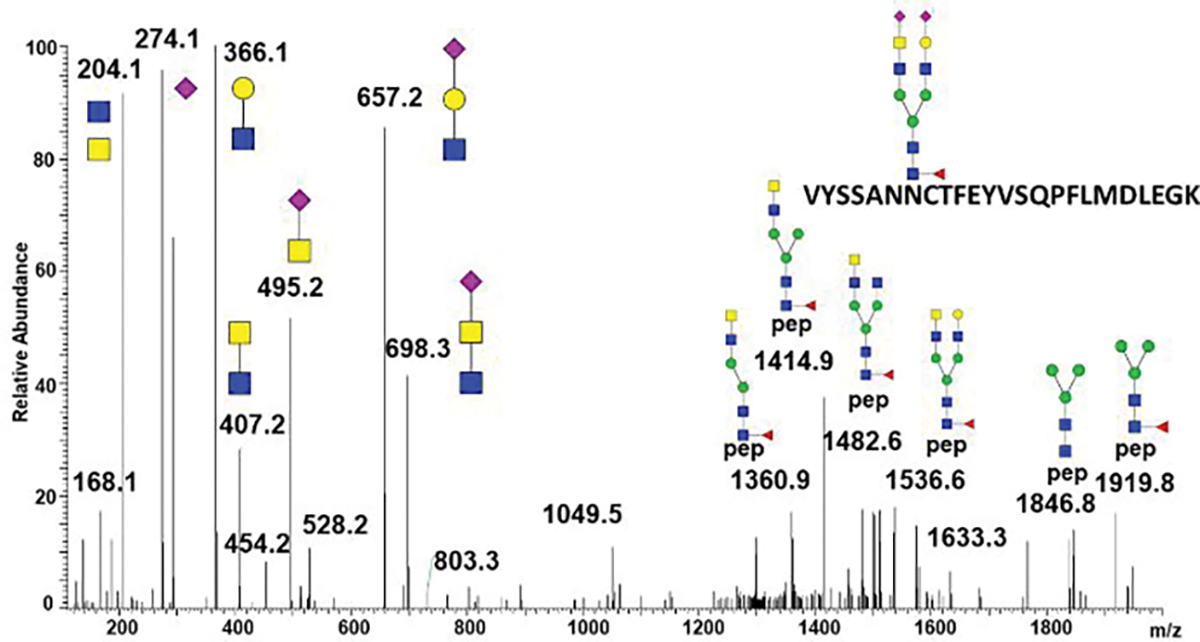
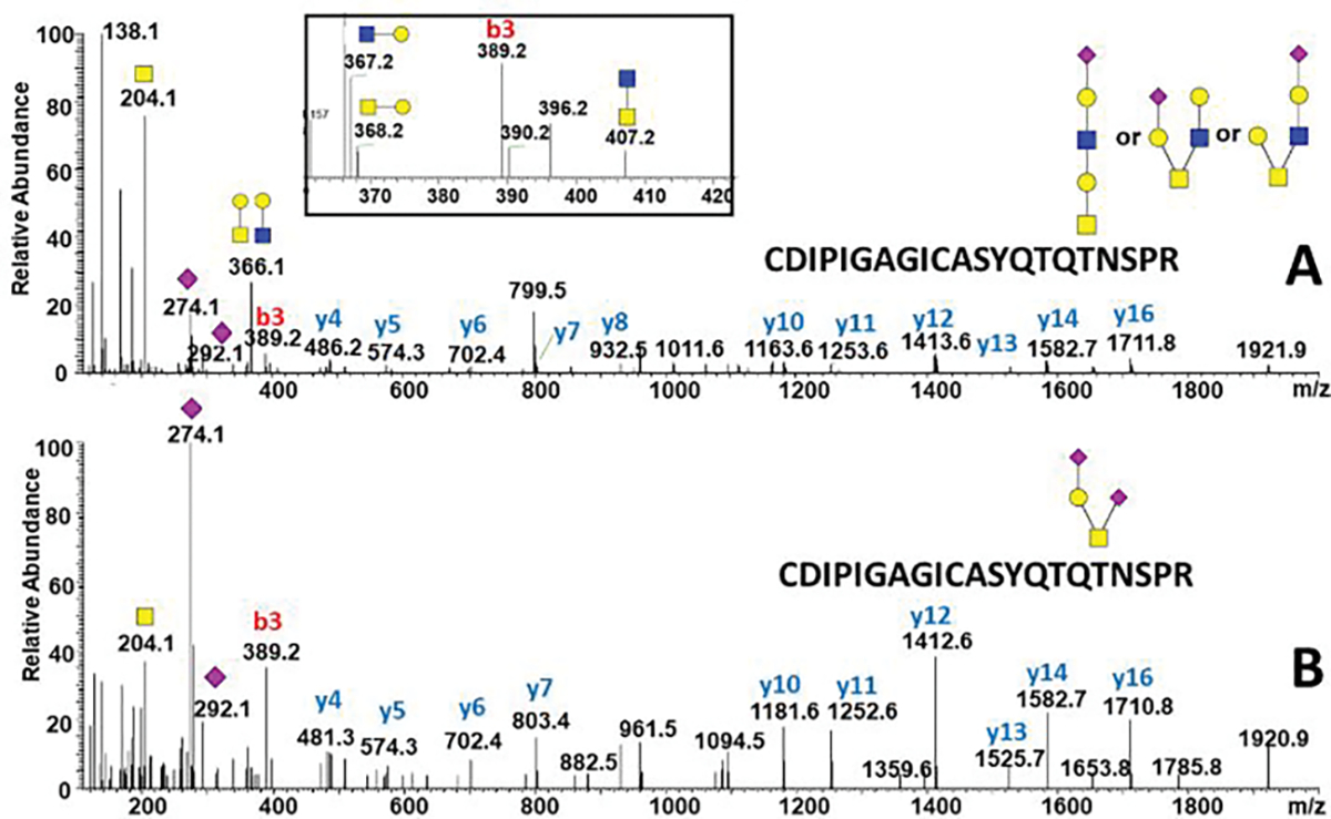
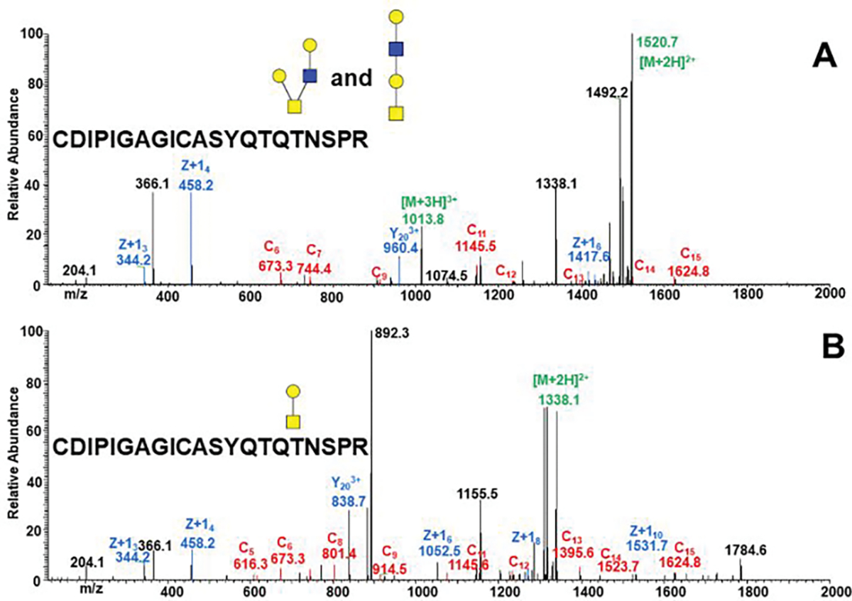
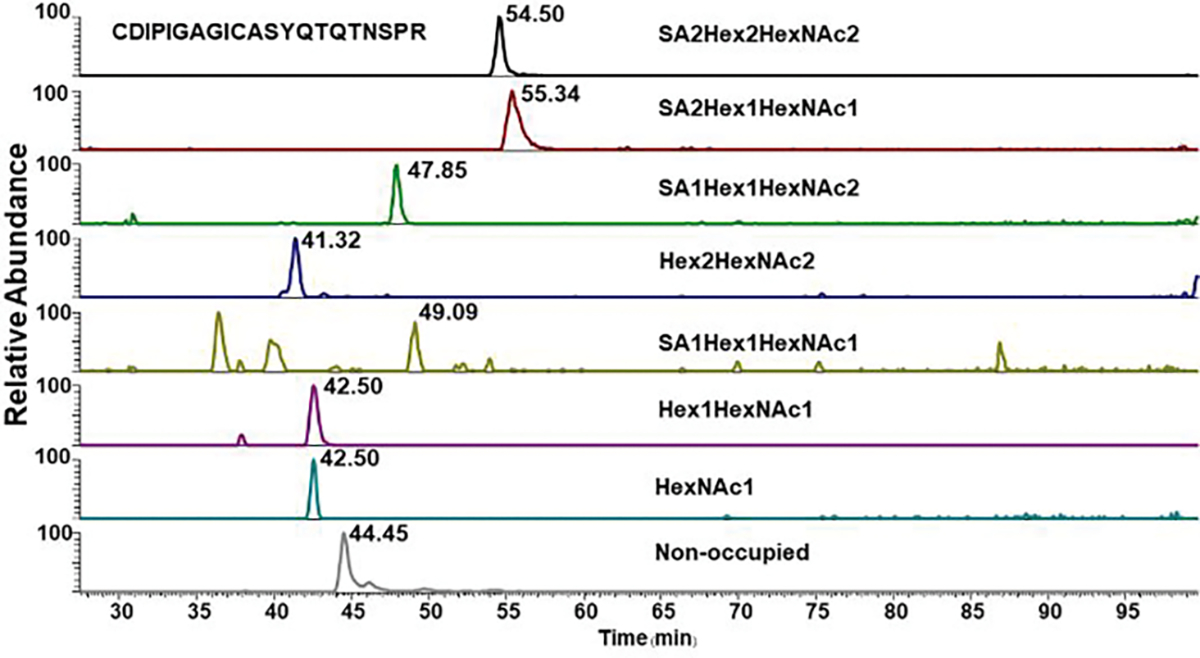
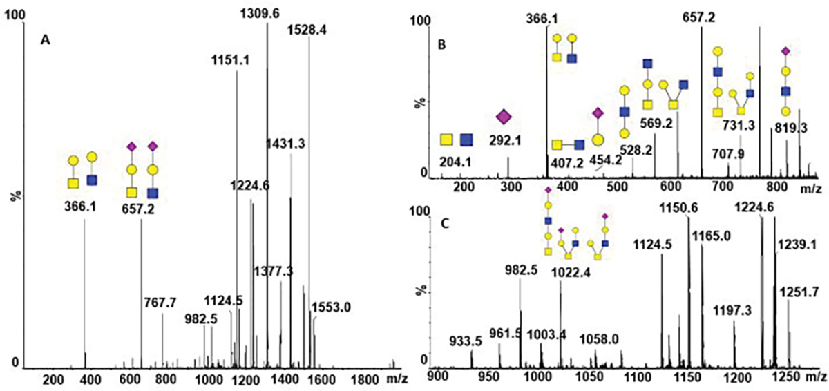
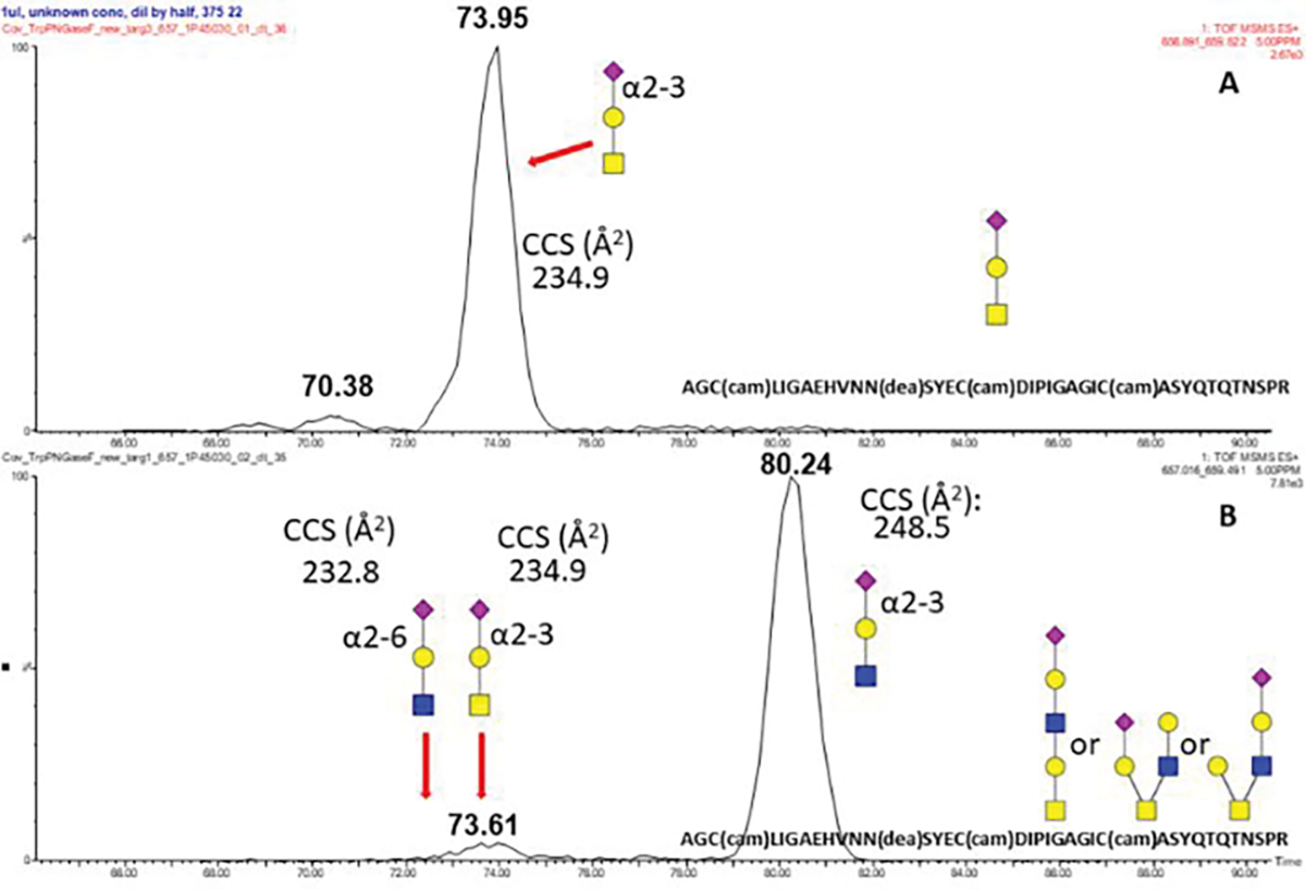
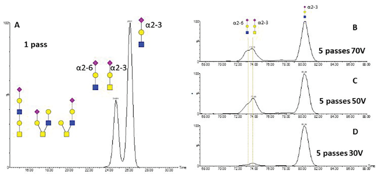
Covid-19 pandemic outbreak is the reason of the current world health crisis. The development of effective antiviral compounds and vaccines requires detailed descriptive studies of SARS-CoV-2 proteins. The SARS-CoV-2 spike (S) protein mediates virion binding to the human cells through its interaction with the ACE2 cell surface receptor and is one of the prime immunization targets. A functional virion is composed of three S1 and three S2 subunits created by furin cleavage of the spike protein at R682, a polybasic cleavage site that differs from the SARS-CoV spike protein of 2002. By analysis of the protein produced in HEK293 cells, we observe that the spike is O-glycosylated on a threonine (T678) near the furin cleavage site occupied by core-1 and core-2 structures. In addition, we have identified eight additional O-glycopeptides on the spike glycoprotein and confirmed that the spike protein is heavily N-glycosylated. Our recently developed liquid chromatography-mass spectrometry methodology allowed us to identify LacdiNAc structural motifs on all occupied N-glycopeptides and polyLacNAc structures on six glycopeptides of the spike protein. In conclusion, our study substantially expands the current knowledge of the spike protein's glycosylation and enables the investigation of the influence of O-glycosylation on its proteolytic activation.


Conflict of interest statement

The authors declare no competing financial interest.

Figures

Figure 1
Figure 1
HCD fragmentation of the N165 glycopeptide carrying an asymmetric biantennary glycan with sialylated LacdiNAc structural motif.
Figure 2.
Figure 2.
HCD tandem mass spectra of the SARS-CoV-2 S protein O-glycosylated on T678 with the following structures: (A) extended core-1 and core-2 structures; (B) disialylated core-1 structure. Inset: oxonium ions in the HCD fragmentation spectrum confirm the presence of core-2 structures.
Figure 3.
Figure 3.
EThcD tandem mass spectra of a tryptic/GluC glycopeptide treated with PNGaseF and non-specific neuraminidase confirms occupancy of the T678 by core-1 (B) and core-2 (A) structures.
Figure 4.
Figure 4.
The CDIPIGAGICASYQTQTNSPR O-glycopeptides of SARS-CoV-2 S protein with the expected glycoform-dependent RT shifts visible in the XIC chromatograms.
Figure 5.
Figure 5.
Beam type tandem mass spectra of the AGC(cam)LIGAEHVNN(dea)SYEC(cam)DIPIGAGIC(cam)ASYQTQTNSPR (SA1Hex2HexNAc2) O-glycopeptide with assigned extended core-1 and core-2 structures. The structures are characterized by the following fragments: (A) oxonium ions 366 and 657, generated from both core-1 and core-2 structures; (B) oxonium ion 407 specific for the core-2 and ions 528 and 819 specific to the extended core-1 structure; and (C) oxonium ion 1022 correspoding to the detached intact glycans.
Figure 6.
Figure 6.
cIMS of the fragment m/z 657 with measured CCS assignments produced by fragmentation of the AGC(cam)LIGAEHVNN(dea)SYEC(cam)DIPIGAGIC(cam)ASYQTQTNSPR (SAHexHexNAc) (A) and (SAHex2HexNAc2) (B) O-glycopeptides produced by tryptic digests and PNGaseF deglycosylation of the SARS-CoV-2 S glycoprotein
Figure 7.
Figure 7.
cIMS of the m/z 657 fragment produced by fragmentation of the AGC(cam)LIGAEHVNN(dea)SYEC(cam)DIPIGAGIC(cam)ASYQTQTNSPR O-Glycopeptide produced by tryptic digest and PNGaseF deglycosylation of the SARS-CoV-2 S glycoprotein using the following settings: (A) one pass cIMS does not resolve SA (2–3) GalGalNAc and SA (2–6)GalGlcNAc; (B). 5 passes cIMS with 70V CE; (C) 5 passes cIMS with 70V CE; and (D) 5 passes cIMS with 70V CE. The ion mobilograms (B,C,D) show that multiple passes improve resolution of isobaric (SA (2–3) GalGalNAc and SA (2–6)GalGlcNAc ) structures and reveals differences in the stability of the sialic acid linkages.

Update of

References

    1. WHO Situation report - 71. Coronavirus Disease 2019 (COVID-19); 2020.
    1. Wu F; Zhao S; Yu B; Chen YM; Wang W; Song ZG; Hu Y; Tao ZW; Tian JH; Pei YY; Yuan ML; Zhang YL; Dai FH; Liu Y; Wang QM; Zheng JJ; Xu L; Holmes EC; Zhang YZ A New Coronavirus Associated with Human Respiratory Disease in China. Nature 2020Mar;579(7798):265–269 - PMC - PubMed
    1. Wrapp D; Wang N; Corbett KS; Goldsmith JA; Hsieh C-L; Abiona O; Graham BS; McLellan JS Cryo-EM Structure of the 2019-NCoV Spike in the Prefusion Conformation. Science (80-. ). 2020. Mar 13;367(6483):1260–1263. - PMC - PubMed
    1. S.A. J; S.M. T; L. G-R; E.M. H; J.E. A; G.J. B; W.D. TJ; L.B. T; M.D. Y; R.J. M; D.M. A; D.E W; J.C. D; K.V. H CD209L (L-SIGN) Is a Receptor for Severe Acute Respiratory Syndrome Coronavirus. Proc Natl Acad Sci U S A. 2004. Nov 2;101(44):15748–53. - PMC - PubMed
    1. Li WH; Moore MJ; Vasilieva N; Sui JH; Wong SK; Berne MA; Somasundaran M; Sullivan JL; Luzuriaga K; Greenough TC; Choe H; Farzan M Single-Cell RNA-Seq Data Analysis on the Receptor ACE2 Expression Reveals the Potential Risk of Different Human Organs Vulnerable to 2019-NCoV Infection. Front Med. 2020. Apr;14(2):185–192 - PMC - PubMed

Publication types

Substances