. 2021 Jan 6;22(1):8.
doi: 10.1186/s12931-020-01598-4.

Resistin-like molecule β acts as a mitogenic factor in hypoxic pulmonary hypertension via the Ca2+-dependent PI3K/Akt/mTOR and PKC/MAPK signaling pathways

Affiliations

Resistin-like molecule β acts as a mitogenic factor in hypoxic pulmonary hypertension via the Ca2+-dependent PI3K/Akt/mTOR and PKC/MAPK signaling pathways

Heshen Tian et al. Respir Res. .

Abstract

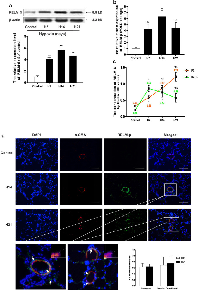
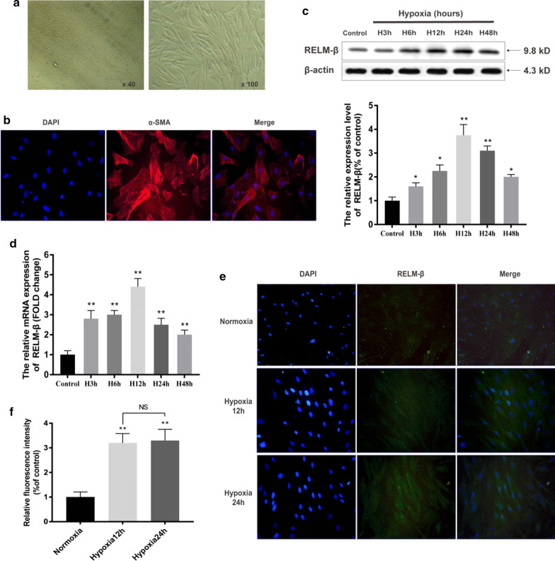
Background: Pulmonary arterial smooth muscle cell (PASMC) proliferation plays a crucial role in hypoxia-induced pulmonary hypertension (HPH). Previous studies have found that resistin-like molecule β (RELM-β) is upregulated de novo in response to hypoxia in cultured human PASMCs (hPASMCs). RELM-β has been reported to promote hPASMC proliferation and is involved in pulmonary vascular remodeling in patients with PAH. However, the expression pattern, effects, and mechanisms of action of RELM-β in HPH remain unclear.

Methods: We assessed the expression pattern, mitogenetic effect, and mechanism of action of RELM-β in a rat HPH model and in hPASMCs.

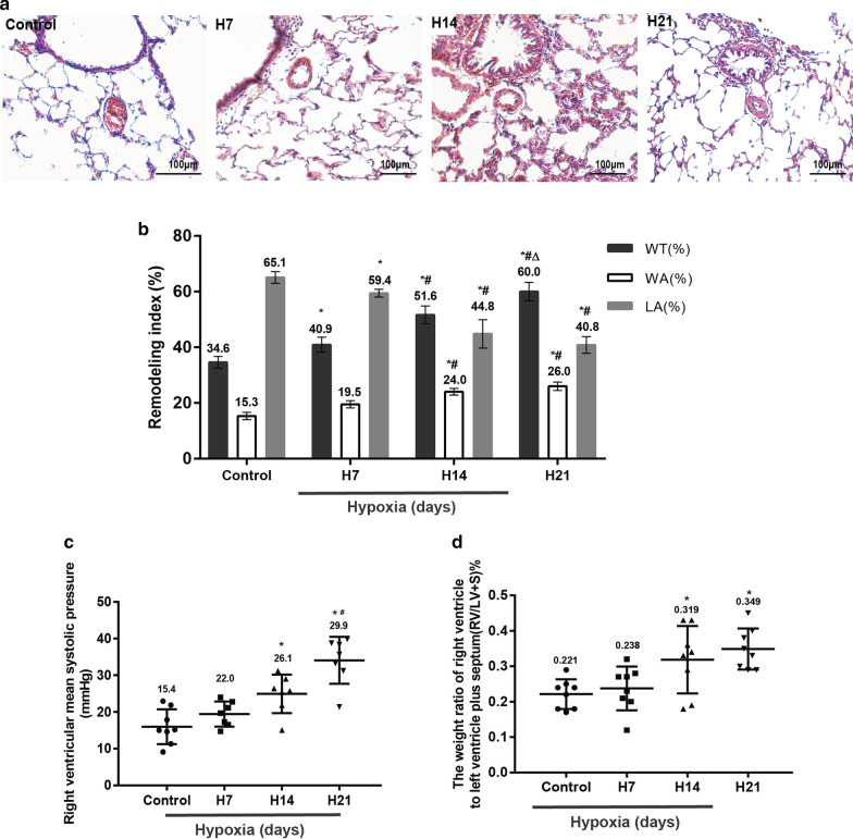
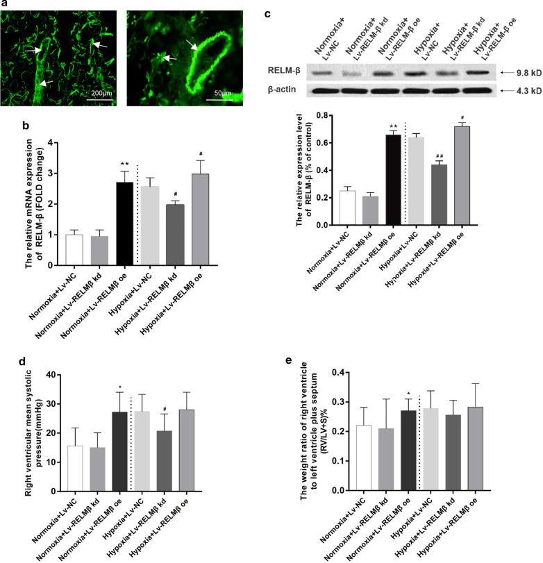
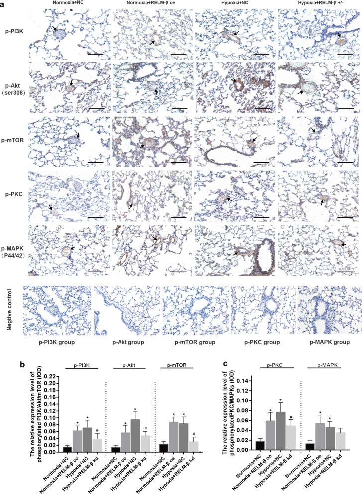
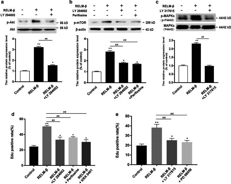
Results: Overexpression of RELM-β caused hemodynamic changes in a rat model of HPH similar to those induced by chronic hypoxia, including increased mean right ventricular systolic pressure (mRVSP), right ventricular hypertrophy index (RVHI) and thickening of small pulmonary arterioles. Knockdown of RELM-β partially blocked the increases in mRVSP, RVHI, and vascular remodeling induced by hypoxia. The phosphorylation levels of the PI3K, Akt, mTOR, PKC, and MAPK proteins were significantly up- or downregulated by RELM-β gene overexpression or silencing, respectively. Recombinant RELM-β protein increased the intracellular Ca2+ concentration in primary cultured hPASMCs and promoted hPASMC proliferation. The mitogenic effects of RELM-β on hPASMCs and the phosphorylation of PI3K, Akt, mTOR, PKC, and MAPK were suppressed by a Ca2+ inhibitor.

Conclusions: Our findings suggest that RELM-β acts as a cytokine-like growth factor in the development of HPH and that the effects of RELM-β are likely to be mediated by the Ca2+-dependent PI3K/Akt/mTOR and PKC/MAPK pathways.

Keywords: Ca2+; Hypoxic pulmonary arterial hypertension; Pulmonary vascular remodeling; Resistin-like molecule β; SOCE; Signaling pathway.


Conflict of interest statement

The authors declare that they have no competing interests.

Figures

Fig. 1
Fig. 1
Verification of the hypoxia-induced rat PAH model. The mean arterial wall thickness at different durations of hypoxia (H&E staining, optical microscope, 400 ×) (a). Morphometric analysis of rat pulmonary arterioles after exposure to normoxia (Control) or hypoxia for 7 days (H7), 14 days (H14) or 21 days (H21) (b). The mRVSP of rats exposed to normoxia or hypoxia for different durations (c). Time course of the RV/LV + S ratio in rats exposed to hypoxia (d). WT(%), ratio of vascular wall thickness to external diameter; LA(%), ratio of lumen area to total vascular area; WA(%), ratio of vascular wall area to total vascular area; n ≥ 6 in each group. *P < 0.05 versus the control group; #P < 0.05 versus group H7; P < 0.05 versus group H14
Fig. 2
Fig. 2
Expression pattern of RELM-β in tissues from an HPH rat model. WB analysis of RELM-β protein expression in rat pulmonary tissue of the control group, H7 group, H14 group and H21 group (a). Quantitative real-time PCR (RT-PCR) results of RELM-β mRNA expression in pulmonary artery muscle at different durations of hypoxia (b). Enzyme-linked immunosorbent assay (ELISA) analysis showing that the serum concentration of RELM-β protein continued to rise, but in the bronchial alveolar lavage fluid, the concentration showed a trend of first increasing and then decreasing, probably due to its paracrine characteristics and regional differences (c). Immunofluorescence colocalization of RELM-β and α-SMA after exposure to hypoxia suggests that RELM-β is expressed in pulmonary arteries (white arrow: pulmonary artery smooth muscle, 400 ×) (d). PB, peripheral blood. n ≥ 6 in each group. *P < 0.05 versus the control group; **P < 0.01 versus the control group; #P < 0.05 versus group H7; P < 0.05 versus group H14
Fig. 3
Fig. 3
Effects of RELM-β on HPH and cardiac and pulmonary artery remodeling. Rat pulmonary tissues were observed over 21 days after intratracheal gene transfer with Lenti6.3-EGFP (1.5 × 108 transducing units in a total volume of 150 μl at a daily dose of 50 μl for 3 days). The transfection efficiency was indicated by the green fluorescence in the lung tissues (the white arrows show that EGFP was abundantly expressed in lung tissues and blood vessels) (a). The relative mRNA level of RELM-β in different treatment groups (b). The WB results of RELM-β and the relative protein expression level of RELM-β in different treatment groups (c). The mean pulmonary arterial pressure of rats in different groups (d). The RV/LV + S ratio in rats in different groups (e). n = 6 rats in each group. *P < 0.05 versus the Normoxia + Lv-NC group; **P < 0.01 versus the Hypoxia + Lv-NC group
Fig. 4
Fig. 4
Proremodeling effect of RELM-β in pulmonary arterioles. The mean arterial wall thickness in different groups was examined by H&E staining (scale bar: 50 μm). Morphometric analysis of rat pulmonary arterioles of different external diameters after exposure to normoxia or hypoxia for 21 days in different groups (a). The walls of small arterioles were stained with antibodies against α-smooth muscle actin and the muscularization of small pulmonary arteries is indicated as NM, PM, and FM in rat lungs 21 days after initial intratracheal instillation of lentivirus. The mean arterial wall thickness examined by H&E staining in different groups (scale bar: 200 μm) (b). NM nonmuscular, PM partially muscular, FM fully muscular. n = 6 rats in each group; the number of vessels counted is ≥ 20 for each bar. *P < 0.05 versus the Normoxia + Lv-NC group; **P < 0.01 versus the Normoxia + Lv-NC group; P < 0.05 versus the Hypoxia + Lv-NC group; △△P < 0.01 versus the Hypoxia + Lv-NC group
Fig. 5
Fig. 5
Alteration of phosphorylated PI3K/Akt/mTOR and PKC/MAPK proteins by RELM-β. Immunohistochemical analysis of phosphorylated PI3K/Akt/mTOR and PKC/MAPK proteins in lung tissue (positive signals in pulmonary arteries were used for analysis); the black arrows indicate that these phosphorylated proteins occur in remolded pulmonary arteries (scale bar: 200 μm) (a). The IOD value of phosphorylated PI3K/Akt/mTOR in different groups (b). The IOD value of phosphorylated PKC/MAPKs in different groups (c). n = 6 rats in each group; the number of vessels counted is ≥ 20 for each bar. *P < 0.05 versus the Normoxia + Lv-NC group. #P < 0.05 versus the Hypoxia + Lv-NC group
Fig. 6
Fig. 6
Alteration of phosphorylated PI3K/Akt/mTOR and PKC/MAPK proteins by RELM-β. The WB results of phosphorylated PI3K/Akt/mTOR and PKC/MAPK protein-isolated pulmonary arteries (a). The relative expression level of p-PI3K in isolated pulmonary arteries (b). The relative expression level of p-Akt in isolated pulmonary arteries (c). The relative expression level of p-mTOR in isolated pulmonary arteries (d). The relative expression level of p-PKC in isolated pulmonary arteries (e). The relative expression level of p-MAPK in isolated pulmonary arteries (f). n = 6 in each group. *P < 0.05 versus the Normoxia + Lv-NC group. **P < 0.01 versus the Normoxia + Lv-NC group; #P < 0.05 versus the Hypoxia + Lv-NC group; ##P < 0.01 versus the Hypoxia + Lv-NC group
Fig. 7
Fig. 7
Hypoxia induces RELM-β expression in primary cultured PASMCs in a time-dependent manner. Light microscopy revealed typical "peak-valley" growth of smooth muscle cells (a). Immunofluorescence staining of α-SMA showed that the cytoplasm of the cells was clearly stained with red fluorescence, which was distributed along the myofilaments and arranged in parallel (b). WB analysis of RELM-β protein expression levels at different durations of hypoxia (3 h, 6 h, 12 h, 24 h, and 48 h) (c). The relative mRNA level of RELM-β at different durations of hypoxia (d). The results in c, d show that RELM-β expression peaked at 12 h and remained high until 48 h of hypoxia. We used immunofluorescence staining of RELM-β to verify this pattern (e, f). n = 6 in each group. *P < 0.05 versus the control group; **P < 0.01 versus the control group
Fig. 8
Fig. 8
RELM-β promotes cell proliferation and Ca2+ release in primary cultured hPASMCs. The cell viability of hPASMCs treated with different concentrations (0 ng/ml, 5 ng/ml, 10 ng/ml, 20 ng/ml, or 40 ng/ml) of RELM-β for 0, 24, 48 or 72 h using the CCK-8 assay (a). The percentage of EdU-positive hPASMCs treated with 0 (control) or 20 ng/ml RELM-β for 48 h using EdU immunofluorescent staining (b, c). RELM-β enhanced the store-operated Ca2+ entry intracellular Ca2+ concentration ([Ca2+]i) in hPASMCs. RELM-β (20 ng/ml for 48 h) enhanced SOCE in hPASMCs. Time course of Δ[Ca2+]i (ΔF340/F380) before and after the restoration of extracellular Ca2+ to 2.5 mM in control hPASMCs (top) and RELM-β-treated (20 ng/ml for 48 h) hPASMCs (bottom) perfused with Ca2+-free Krebs–Ringer bicarbonate solution (KRS) containing 10 μM cyclopiazonic acid (CPA) and 5 μM nifedipine. Ca2+-free KRS also contained 1 mM EGTA to chelate any residual Ca2+. The average peak change in Δ[Ca2+]i (ΔF340/F380) after restoration of extracellular Ca2+(SOCE) in control and RELM-β-treated hPASMCs (right) (d). n = 3 in each group. *P < 0.05 versus control group; **P < 0.01 versus control group
Fig. 9
Fig. 9
Ca2+ is involved in the activation of the PI3K/Akt/mTOR and PKC/MAPK pathways and PASMC proliferation by RELM-β stimulation. The WB results and relative p-PI3K, p-Akt, and p-mTOR protein levels of PASMCs treated with 0 (control) or 20 ng/ml RELM-β, RELM-β + 10 μM BAPTA-AM, or RELM-β + 4 mM EGTA (with 0 mM extracellular Ca2+) (a). The WB results and relative p-PKC and p-MAPK protein levels of PASMCs treated with 0 (control) or 20 ng/ml RELM-β, RELM-β + BAPTA, or RELM-β + EGTA for 48 h (b). The percentage of EdU-positive PASMCs treated with 0 (control) or 20 ng/ml RELM-β, RELM-β + BAPTA-AM, or RELM-β + EGTA for 48 h using EdU immunofluorescent staining (c). n = 3 in each group. *P < 0.05 versus the control group; #P < 0.05 versus the RELM-β group
Fig. 10
Fig. 10
Involvement of PI3K/Akt/mTOR and PKC/MAPKs in RELM-β-induced PASMC proliferation. The WB results and relative p-Akt protein levels of PASMCs treated with 0 (control) or 20 ng/ml RELM-β or RELM-β + LY294002 (10 μM, a PI3K inhibitor) (a). The WB results and relative p-mTOR protein levels of PASMCs treated with 0 (control) or 20 ng/ml RELM-β, RELM-β + LY294002 and RELM-β + perifosine (100 nM, an Akt inhibitor) (b). The WB results and relative p-MAPK protein levels of PASMCs treated with 0 (control) or 20 ng/ml RELM-β or RELM-β + LY317615 (10 μM, a PKC inhibitor) (c). The percentage of EdU-positive PASMCs treated with 0 (control) or 20 ng/ml RELM-β, RELM-β + LY294002, RELM-β + perifosine, or RELM-β + rapamycin (100 nM, a mTOR inhibitor) (d) and with RELM-β + LY317615 or RELM-β + PD98059 (20 μM, a MAPK inhibitor) (e) for 48 h using EdU immunofluorescence staining (c). n = 3 in each group. *P < 0.05 versus the control group; **P < 0.01 versus the control group; ##P < 0.01 between two groups indicated by a line

References

    1. Stenmark KR, Fagan KA, Frid MG. Hypoxia-induced pulmonary vascular remodeling: cellular and molecular mechanisms. Circ Res. 2006;99(7):675–691. doi: 10.1161/01.RES.0000243584.45145.3f. - DOI - PubMed
    1. Sahay S. Evaluation and classification of pulmonary arterial hypertension. J Thorac Dis. 2019;11(Suppl 14):S1789–S1799. doi: 10.21037/jtd.2019.08.54. - DOI - PMC - PubMed
    1. Opitz I, Ulrich S. Pulmonary hypertension in chronic obstructive pulmonary disease and emphysema patients: prevalence, therapeutic options and pulmonary circulatory effects of lung volume reduction surgery. J Thorac Dis. 2018;10(Suppl 23):S2763–S2774. doi: 10.21037/jtd.2018.07.63. - DOI - PMC - PubMed
    1. Campo A, Mathai SC, Le Pavec J, et al. Outcomes of hospitalisation for right heart failure in pulmonary arterial hypertension. Eur Respir J. 2011;38(2):359–367. doi: 10.1183/09031936.00148310. - DOI - PubMed
    1. Thenappan T, Prins KW, Pritzker MR, Scandurra J, Volmers K, Weir EK. The critical role of pulmonary arterial compliance in pulmonary hypertension. Ann Am Thorac Soc. 2016;13(2):276–284. - PMC - PubMed

MeSH terms

LinkOut - more resources