

Molecular detection and characterisation of Domestic Cat Hepadnavirus (DCH) from blood and liver tissues of cats in Malaysia
- PMID: 33407487
- PMCID: PMC7788742
- DOI: 10.1186/s12917-020-02700-0
Molecular detection and characterisation of Domestic Cat Hepadnavirus (DCH) from blood and liver tissues of cats in Malaysia
Abstract
Background: A new domestic cat hepadnavirus (DCH, family Hepadnaviridae) was first reported from whole blood samples of domestic cats in Australia in 2018, and from cat serum samples in Italy in 2019. The pathogenesis of DCH is unknown, but it was reported in cats with viraemia (6.5-10.8%), chronic hepatitis (43%) and hepatocellular carcinoma (28%). Recent reports suggest that DCH resembles the human hepatitis B virus (HBV) and its related hepatopathies. This study aims to detect and characterize DCH among domestic cats in Malaysia. A cross-sectional study was performed on 253 cats, of which 87 had paired blood and liver samples, entailing whole-genome sequencing and phylogenetic analysis of DCH from a liver tissue sample.
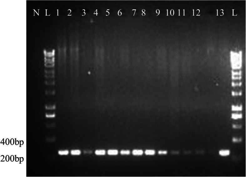
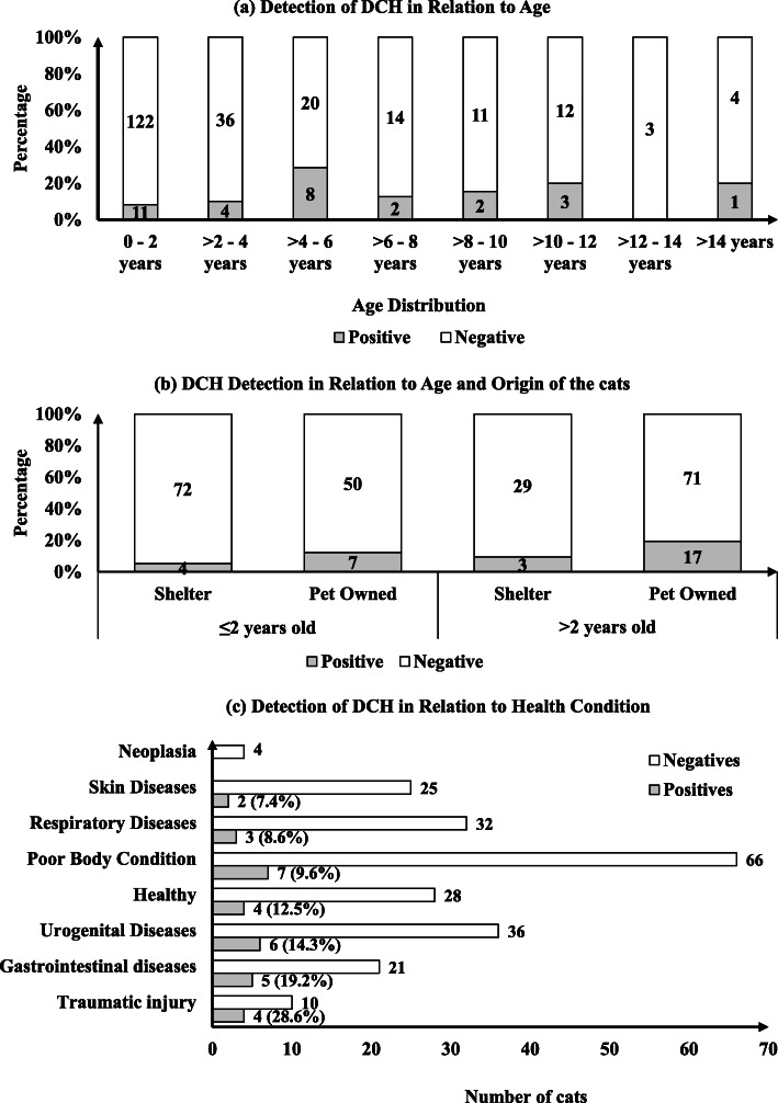
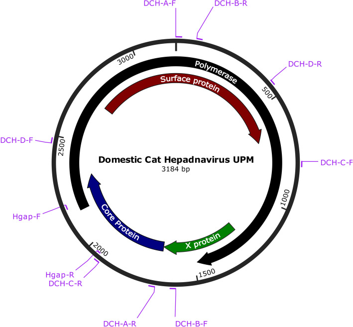
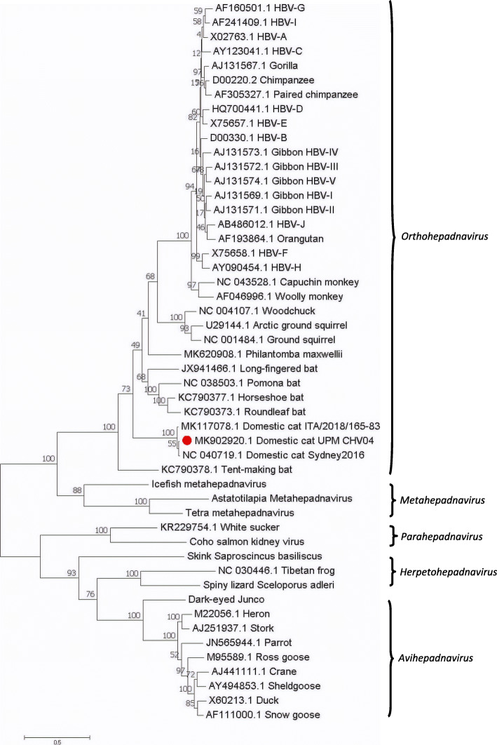
Results: Among the 253 cats included in this study, 12.3% of the whole blood samples tested positive for DCH. The detection rate was significantly higher in pet cats (16.6%, n = 24/145) compared to shelter cats (6.5%, n = 7/108). Liver tissues showed higher a DCH detection rate (14.9%, n = 13/87) compared to blood; 5 out of these 13 cats tested positive for DCH in their paired liver and blood samples. Serum alanine transaminase (ALT) was elevated (> 95 units/L) in 12 out of the 23 DCH-positive cats (52.2%, p = 0.012). Whole-genome sequence analysis revealed that the Malaysian DCH strain, with a genome size of 3184 bp, had 98.3% and 97.5% nucleotide identities to the Australian and Italian strains, respectively. The phylogenetic analysis demonstrated that the Malaysian DCH genome was clustered closely to the Australian strain, suggesting that they belong to the same geographically-determined genetic pool (Australasia).
Conclusions: This study provided insights into a Malaysian DCH strain that was detected from a liver tissue. Interestingly, pet cats or cats with elevated ALT were significantly more likely to be DCH positive. Cats with positive DCH detection from liver tissues may not necessarily have viraemia. The impact of this virus on inducing liver diseases in felines warrants further investigation.
Keywords: Clinical pathology; Feline; Hepadnavirus; Liver; Malaysia; PCR; Phylogenetic analysis; Prevalence; Risk factors.
Conflict of interest statement
The authors declare that they have no competing interests.
Figures
References
-
- Urban S, Schulze A, Dandri M, Petersen J. The replication cycle of hepatitis B virus. J Hepatol. 2010;52(2):282–4. - PubMed
-
- Monjardino J. Hepadnaviridae. Virus Taxonomy. 2012;445–455.
MeSH terms
Substances
Grants and funding
LinkOut - more resources
Full Text Sources
Other Literature Sources
Miscellaneous
