Review
. 2021 Jan 7;19(1):4.
doi: 10.1186/s12958-020-00686-w.

FSH regulates RA signaling to commit spermatogonia into differentiation pathway and meiosis

Affiliations
Review

FSH regulates RA signaling to commit spermatogonia into differentiation pathway and meiosis

Maryam Khanehzad et al. Reprod Biol Endocrinol. .

Abstract

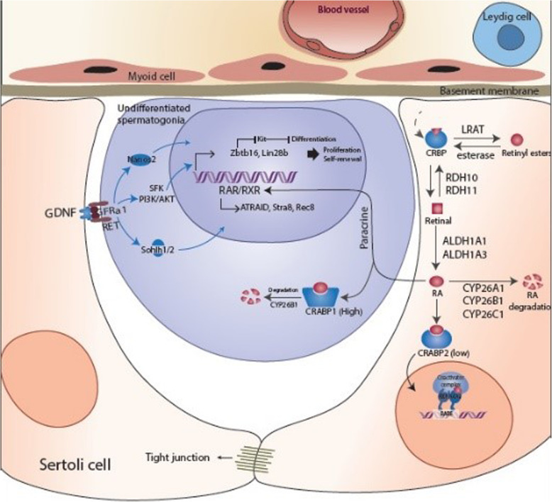
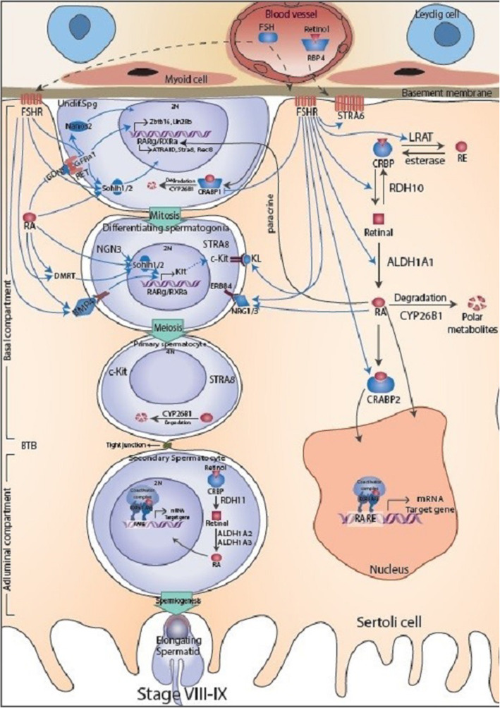
Background: Spermatogenesis is a complex process that is controlled by interactions between germ cells and somatic cells. The commitment of undifferentiated spermatogonia to differentiating spermatogonia and normal spermatogenesis requires the action of gonadotropins. Additionally, numerous studies revealed the role of retinoic acid signaling in induction of germ cell differentiation and meiosis entry.

Main text: Recent studies have shown that expression of several RA signaling molecules including Rdh10, Aldh1a2, Crabp1/2 are influenced by changes in gonadotropin levels. Components of signaling pathways that are regulated by FSH signaling such as GDNF, Sohlh1/2, c-Kit, DMRT, BMP4 and NRGs along with transcription factors that are important for proliferation and differentiation of spermatogonia are also affected by retinoic acid signaling.

Conclusion: According to all studies that demonstrate the interface between FSH and RA signaling, we suggest that RA may trigger spermatogonia differentiation and initiation of meiosis through regulation by FSH signaling in testis. Therefore, to the best of our knowledge, this is the first time that the correlation between FSH and RA signaling in spermatogenesis is highlighted.

Keywords: Differentiation; FSH; Retinoic acid; Spermatogenesis; Spermatogonia.


Conflict of interest statement

The authors declare that they have no competing interests.

Figures

Fig. 1
Fig. 1
The possible paracrine mechanisms controlling self-renewal of spermatogonia in juvenile testis. Spermatogonia are in close contact with Sertoli cells which provide structural support and paracrine factors to the developing spermatogenic cells. The self-renewal and proliferation factor GDNF expressed by Sertoli cells is responsible for transcriptional activation of Zbtb16 and Lin28B and thereby preventing spermatogonia to enter differentiation pathway. Sertoli cells are the source of paracrine RA signaling pathway. In Sertoli cells, retinol is oxidized to retinal by RDH10 and then to RA by RALDH1a1. However, the amount of RA produced by Sertoli cells within juvenile testis is not robust to initiate spermatogonia differentiation possibly due to the low amount of CRABP2 which is considered for activating of RA signaling in Sertoli cells as well as the high amount of CRABP which is considered for degrading of RA signaling within spermatogonia. Black arrows refer to retinol metabolism and RA signaling pathway. Blue arrows refer to endogenous factors that promote spermatogonia proliferation and self-renewal but at the same time inhibits spermatogonial differentiation and meiotic entry. RBP4, retinol binding protein 4; STRA6, stimulated by retinoic acid gene 6 cell membrane receptor; CRBP, cellular retinol binding protein; LRAT, lecithin retinol transferase; RDH10, retinol dehydrogenase 10; RALDH1a1, retinaldehyde dehydrogenase 1a1; RA, retinoic acid; CYP26, cytochrome P-450 enzymes from the cyp26 family; CRABP, cellular retinoic acid binding protein; RAR, retinoic acid receptor; RXR, rexinoid receptor;. RARE, retinoic acid respond element; ATRAID, All-Trans Retinoic Acid Induced Differentiation Factor; Stra8, stimulated by retinoic acid 8; Rec8, REC8 like meiotic recombination protein; GDNF, Glial cell-derived neurotrophic factor; GFRα1, GDNF family receptor α1; Nanos2, Nanos Homolog 2; Sohlh1/2, Spermatogenesis- And Oogenesis-Specific Basic Helix-Loop-Helix-Containing Protein 1,2; Zbtb16, Zinc finger and BTB domain containing 16; Lin28B, Lin-28 homolog B; c-Kit, V-Kit Hardy-Zuckerman 4 Feline Sarcoma Viral Oncogene-Like Protein
Fig. 2
Fig. 2
The possible paracrine mechanisms controlling differentiation of spermatogonia in adult testis. The spermatogenic cells arise from differentiation of undifferentiated type A spermatogonia (2n). The Ap spermatogonia can divide to form an Ap and a differentiatimg type B spermatogonia (2n) through the cycle of the seminiferous epithelium stages VIII to IX. B spermatogonia divide randomly and differentiate to form primary (4n) and secondary spermatocytes (2n) through meiotic divisions. The secondary spermatocytes proceed through the rest of meiosis to form elongating spermatids (n). Differentiation of spermatogonia occurs during stages of VII-VIII when the RA concentration is at the highest levels. FSH is necessary for early-stage of spermatogonia differentiation and their meiotic entry. FSH induces spermatogonia differentiation and their meiotic entry thorough controlling RA signaling and several growth factors acting on the early stages of mammalian spermatogenesis. In one hand, FSH probably induces the activation of RA signaling pathway notably through increase Rdh10, Aldh1a1, Crabp2 in Sertoli cells and decrease Crabp1 in spermatogonia and thereby provide a robust paracrine factor necessary for induction of spermatogonia differentiation. In another hand, FSH induces components of signaling pathways regulated by RA signaling such as Sohlh1/2, Kit ligand, DMRT, BMP4 and NRGs along with transcription factors that are important for proliferation and differentiation of spermatogonia and their meiotic entry. Exogenous and endogenous factors are represented in Sertoli and germ cells, respectively. Black arrows refer to retinol metabolism and RA signaling pathway. The succession of the various types of germ cells during the stages of VIII-IX seminiferous epithelium is represented between Sertoli cells: AS, undifferentiated type A spermatogonia; BS, differentiating type B spermatogonia; PS, pre-leptotene or primary spermatocytes; SS, secondary spermatocytes; ES, elongating spermatocytes. FSH, Follicle Stimulating Hormone, FSHR, Follicle Stimulating Hormone receptor; KL, Kit ligand ligand; DMRT, Doublesex And Mab-3 Related Transcription Factor; BMP4, Bone Morphogenetic Protein 4; NRG, Neuregulin; BTB, blood testis barrier

References

    1. Plant TM. Undifferentiated primate spermatogonia and their endocrine control. Trends Endocrinol Metab. 2010;21(8):488–495. doi: 10.1016/j.tem.2010.03.001. - DOI - PMC - PubMed
    1. Feng CW, Bowles J, Koopman P. Control of mammalian germ cell entry into meiosis. Mol Cell Endocrinol. 2014;382:488–497. doi: 10.1016/j.mce.2013.09.026. - DOI - PubMed
    1. Plant TM, Marshall GR. The functional significance of FSH in spermatogenesis and the control of its secretion in male primates. Endocr Rev. 2001;22:764–786. doi: 10.1210/edrv.22.6.0446. - DOI - PubMed
    1. Plant TM, Witchel SF. Puberty in nonhuman primates and humans. In: Neill JD, editor. The physiology of reproduction. San Diego: Academic Press/Elsevier; 2006. pp. 2177–2230.
    1. Plant TM, Ramaswamy S, Simorangkir DR, Marshall GR. Postnatal and pubertal development of the rhesus monkey (Macaca mulatta) testis. Ann N Y Acad Sci. 2005;1061:149–162. doi: 10.1196/annals.1336.016. - DOI - PubMed

LinkOut - more resources