Comparative Study
. 2021 Jan 6;18(1):10.
doi: 10.1186/s12974-020-02054-2.

Comparisons of neuroinflammation, microglial activation, and degeneration of the locus coeruleus-norepinephrine system in APP/PS1 and aging mice

Affiliations
Comparative Study

Comparisons of neuroinflammation, microglial activation, and degeneration of the locus coeruleus-norepinephrine system in APP/PS1 and aging mice

Song Cao et al. J Neuroinflammation. .

Abstract

Background: The role of microglia in Alzheimer's disease (AD) pathogenesis is becoming increasingly important, as activation of these cell types likely contributes to both pathological and protective processes associated with all phases of the disease. During early AD pathogenesis, one of the first areas of degeneration is the locus coeruleus (LC), which provides broad innervation of the central nervous system and facilitates norepinephrine (NE) transmission. Though the LC-NE is likely to influence microglial dynamics, it is unclear how these systems change with AD compared to otherwise healthy aging.

Methods: In this study, we evaluated the dynamic changes of neuroinflammation and neurodegeneration in the LC-NE system in the brain and spinal cord of APP/PS1 mice and aged WT mice using immunofluorescence and ELISA.

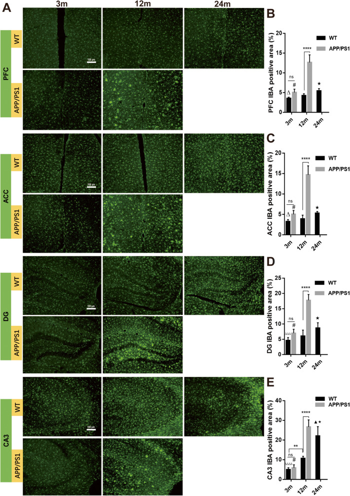
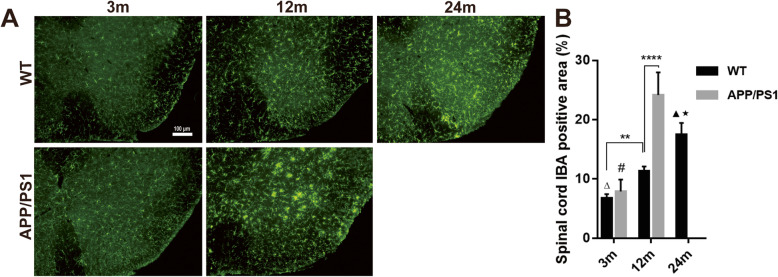
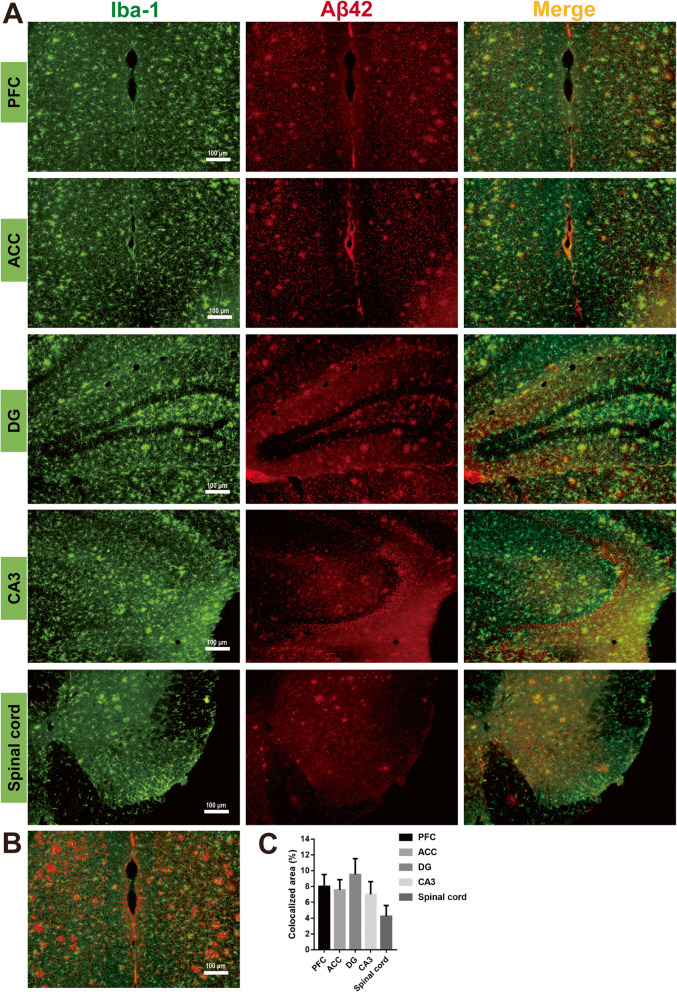
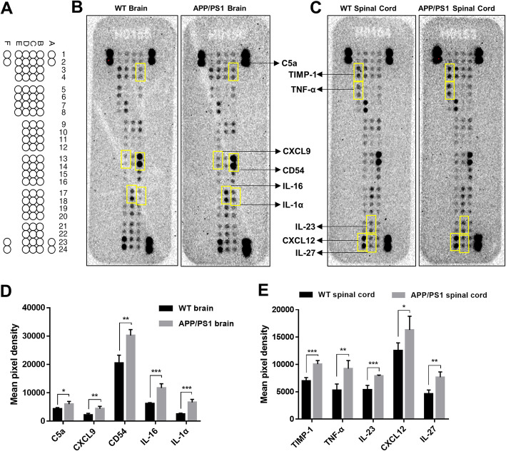
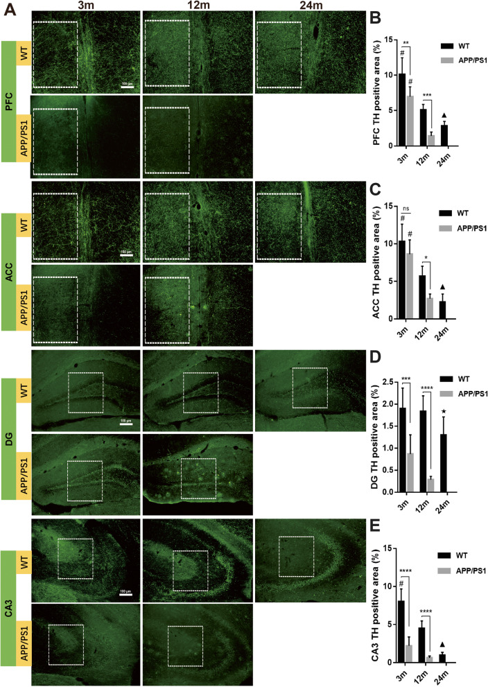
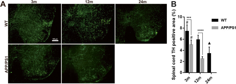
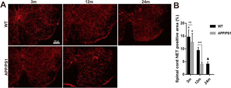
Results: Our results demonstrated increased expression of inflammatory cytokines and microglial activation observed in the cortex, hippocampus, and spinal cord of APP/PS1 compared to WT mice. LC-NE neuron and fiber loss as well as reduced norepinephrine transporter (NET) expression was more evident in APP/PS1 mice, although NE levels were similar between 12-month-old APP/PS1 and WT mice. Notably, the degree of microglial activation, LC-NE nerve fiber loss, and NET reduction in the brain and spinal cord were more severe in 12-month-old APP/PS1 compared to 12- and 24-month-old WT mice.

Conclusion: These results suggest that elevated neuroinflammation and microglial activation in the brain and spinal cord of APP/PS1 mice correlate with significant degeneration of the LC-NE system.

Keywords: Alzheimer’s disease; Dopaminergic; Locus coeruleus; Microglia; Noradrenergic; Norepinephrine transporter; Norepinephrine/noradrenaline; Spinal cord.


Conflict of interest statement

The authors declare that they have no competing interests.

Figures

Fig. 1
Fig. 1
Microglia immunofluorescence of different brain areas in APP/PS1 and WT mice. a Immunofluorescence demonstrated that the Iba-1+ area in the b medial prefrontal cortex (PFC), c anterior cingulate cortex, and d hippocampal DG, and e CA3 increased significantly with aging in WT and APP/PS1 mice. In 12-month-old APP/PS1 mice, Iba-1 expression was significantly more than that of 12-month-old WT mice and 3-month-old APP/PS1 mice. In addition, in PFC, ACC, and DG, the Iba-1+ area in 12-month-old APP/PS1 mice was significantly more than in 24-month old WT mice. n = 6. ns, not significant. **P < 0.01. ****P < 0.0001. #P < 0.0001 compared with 12-month-old APP/PS1 mice. ▲P < 0.05 compared with 12-month-old WT mice. ★P < 0.0001 compared with 12-month-old APP/PS1 mice. ΔP < 0.05 compared with 24-month-old WT mice. ΔΔΔP < 0.001 compared with 24-month-old WT mice. ΔΔΔΔP < 0.0001 compared with 24-month-old WT mice
Fig. 2
Fig. 2
Microglia immunofluorescence in the spinal cord of APP/PS1 and WT mice. a Iba-1 expression in the lumbar spinal cord was detected by immunofluorescence. b Iba-1 expression in the two groups of mice increased significantly with aging. Iba-1+ area in 12-month-old APP/PS1 mice was significantly more than that of 12-month-old WT mice and 3-month-old APP/PS1 mice; it was even more than that in 24-month-old WT mice. n = 6. ns, not significant. **P < 0.01, ****P < 0.0001, #P < 0.0001 compared with 12-month-old APP/PS1 mice. ΔP < 0.0001 compared with 24-month-old WT mice. ▲P < 0.001 compared with 12 m WT mice. ★P < 0.0001 compared with 12 m APP/PS1 mice.
Fig. 3
Fig. 3
Microglia accumulating around Aβ42+ plaques in the brain and spinal cord. a Iba-1 and Aβ42 co-staining in PFC, ACC, DG, CA3, and lumbar spinal cord from 12-month-old APP/PS1 mice showed that a large number of microglia activated and accumulated around the Aβ42+ plaques in the brain and in the grey matter of the spinal cord. b, c Iba-1 and Aβ42 colocalized areas (yellow color in a) in PFC were selected and quantified with Image J
Fig. 4
Fig. 4
Comparisons of cytokine expression in the brain and spinal cord of WT and APP/PS1 mice. a Cytokine array coordinates on membranes. b, c Representative membrane images of cytokine expression in the b brain and c spinal cord. Compared with WT mice, d 5 cytokines were significantly upregulated in the brain of APP/PS1 mice, and e 5 cytokines were significantly upregulated in the spinal cord of APP/PS1 mice. n = 4 in each group. Statistical analyses were conducted with multiple t tests with false discovery rate (FDR) correction. *P < 0.05, **P < 0.01. ***P < 0.001
Fig. 5
Fig. 5
Immunofluorescence of LC-NE neuron and NE detection in the brain and spinal cord. a TH staining shows TH+ LC-NE neurons and fibers. b TH+ neurons were counted manually, and data indicated that LC-NE neurons in WT mice are gradually lost with aging, although the neuron number of 24-month-old WT mice was not significantly less than that of 3-month-old and 12-month-old WT mice. In 12-month-old APP/PS1 mice, this number decreased significantly compared to that of the 12-month-old WT mice. Three brain slices from each mouse were analyzed and averaged. n = 6. ns, not significant. **P < 0.01. #P < 0.01 compared with 12-month-old APP/PS1 mice. 4V, fourth ventricle. c NE level in the brain and spinal cord of APP/PS1 and WT mice was detected with ELISA. No difference was detected in brain or spinal cord between groups. n = 6, ns, not significant
Fig. 6
Fig. 6
Detection of TH+ nerve fibers in the brains of APP/PS1 and WT mice. a Immunofluorescence detection of TH+ nerve fibers in different brain regions showed that in the b medial prefrontal cortex, c anterior cingulate, d DG, and e CA3, TH+ nerve fibers gradually decreased with aging. In 12-month-old APP/PS1 mice, TH+ area was significantly reduced compared with WT mice of the same age and 3-month-old APP/PS1 mice. TH+ area in the brain slices of 3-month-old and 12-month-old APP/PS1 mice was significantly lower than that of WT mice of the same age. In DG, TH+ area of 12-month-old APP/PS1 mice was even significantly lower than that of 24-month-old WT mice. White box indicated region of interests used for the calculation of TH+ area. n = 6. ns, not significant. *P < 0.05, **P < 0.01, ***P < 0.001, ****P < 0.0001, #P < 0.05 compared with other time points in the same group. ▲P < 0.05 compared with 12 m WT mice. ★P < 0.01 compared with 12 m APP/PS1 mice
Fig. 7
Fig. 7
Detection of TH+ nerve fibers in spinal cord of APP/PS1 and WT mice. a TH+ nerve fibers in the lumbar spinal cord were detected by immunofluorescence. b TH+ fibers in the two groups of mice decreased with aging. The percentage of TH+ area in 3-month-old and 12-month-old APP/PS1 mice was significantly lower than that of WT mice of the same age. The TH expression in 12-month-old APP/PS1 mice was significantly less than that of 3-month-old APP/PS1 mice, and it was even less than that of 24-month-old WT mice. n = 6. ***P < 0.001, ****P < 0.0001, #P < 0.05 compared with other time points in the same group. ▲P < 0.05 compared with 12 m WT mice
Fig. 8
Fig. 8
Detection of NET+ nerve fibers in APP/PS1 and WT mice. a Immunofluorescence detected NET+ nerve fibers in b medial prefrontal cortex, c anterior cingulate cortex, d DG, and e CA3. In these areas, NET+ nerve fibers gradually decreased with aging. NET+ nerve fibers in 12-month-old APP/PS1 mice were significantly less compared with WT mice of the same age and 3-month-old APP/PS1 mice. In ACC, DG, and CA3 of 12-month-old APP/PS1 mice, the NET+ area was less than that of 24-month-old WT mice. n = 6. Comparisons between APP/PS1 mice and WT mice and comparisons among 3 WT groups were estimated with two-way ANOVA and followed by Tukey’s post hoc multiple comparison tests. ns, not significant. **P < 0.01, ***P < 0.001, ****P < 0.0001, #P < 0.05 compared with other time points in the same group. ▲P < 0.05 compared with 12-month-old WT mice. ★P < 0.01 compared with 12-month-old APP/PS1 mice
Fig. 9
Fig. 9
Spinal NET+ nerve fiber detection in APP/PS1 and WT mice. a The NET+ nerve fibers in the lumbar spinal cord were detected by immunofluorescence. b The NET+ area in the two groups of mice decreased significantly with aging. The expression of NET in 12-month-old APP/PS1 mice was significantly lower than those of WT mice of the same age and 3-month-old APP/PS1 mice. n = 6. ns, not significant. ***P < 0.001, #P < 0.05 compared with other time points in the same group. ▲P < 0.001 compared with 12 m WT mice

Similar articles

Cited by

References

    1. Chakraborty S, Lennon JC, Malkaram SA, Zeng Y, Fisher DW, Dong H. Serotonergic system, cognition, and BPSD in Alzheimer’s disease. Neurosci Lett. 2019;704:36–44. doi: 10.1016/j.neulet.2019.03.050. - DOI - PMC - PubMed
    1. Cao S, Fisher DW, Yu T, Dong H. The link between chronic pain and Alzheimer’s disease. J Neuroinflammation. 2019;16:204. doi: 10.1186/s12974-019-1608-z. - DOI - PMC - PubMed
    1. Vossel KA, Tartaglia MC, Nygaard HB, Zeman AZ, Miller BL. Epileptic activity in Alzheimer’s disease: causes and clinical relevance. Lancet Neurol. 2017;16:311–322. doi: 10.1016/S1474-4422(17)30044-3. - DOI - PMC - PubMed
    1. Stubbs B, Thompson T, Solmi M, Vancampfort D, Sergi G, Luchini C, Veronese N. Is pain sensitivity altered in people with Alzheimer’s disease? A systematic review and meta-analysis of experimental pain research. Exp Gerontol. 2016;82:30–38. doi: 10.1016/j.exger.2016.05.016. - DOI - PubMed
    1. Jensen-Dahm C, Werner MU, Dahl JB, Jensen TS, Ballegaard M, Hejl AM, Waldemar G. Quantitative sensory testing and pain tolerance in patients with mild to moderate Alzheimer disease compared to healthy control subjects. Pain. 2014;155:1439–1445. doi: 10.1016/j.pain.2013.12.031. - DOI - PubMed

Publication types

MeSH terms

LinkOut - more resources