

Shrinking lung syndrome treated with rituximab in pediatric systemic lupus erythematosus: a case report and review of the literature
- PMID: 33407629
- PMCID: PMC7789161
- DOI: 10.1186/s12969-020-00491-0
Shrinking lung syndrome treated with rituximab in pediatric systemic lupus erythematosus: a case report and review of the literature
Abstract
Background: Shrinking lung syndrome (SLS), a rare complication of systemic lupus erythematosus (SLE) characterized by dyspnea, low lung volumes, and a restrictive pattern on pulmonary function tests (PFTs), has only been reported in a few children. Given the rarity of SLS there is a paucity of literature regarding its optimal treatment. Outcomes are variable, with case reports documenting some improvement in most patients treated with corticosteroids, with or without additional immunosuppressive agents. However, most reported patients did not recover normal lung function. We report full recovery of a child with SLE and SLS following treatment with rituximab and review the current literature.
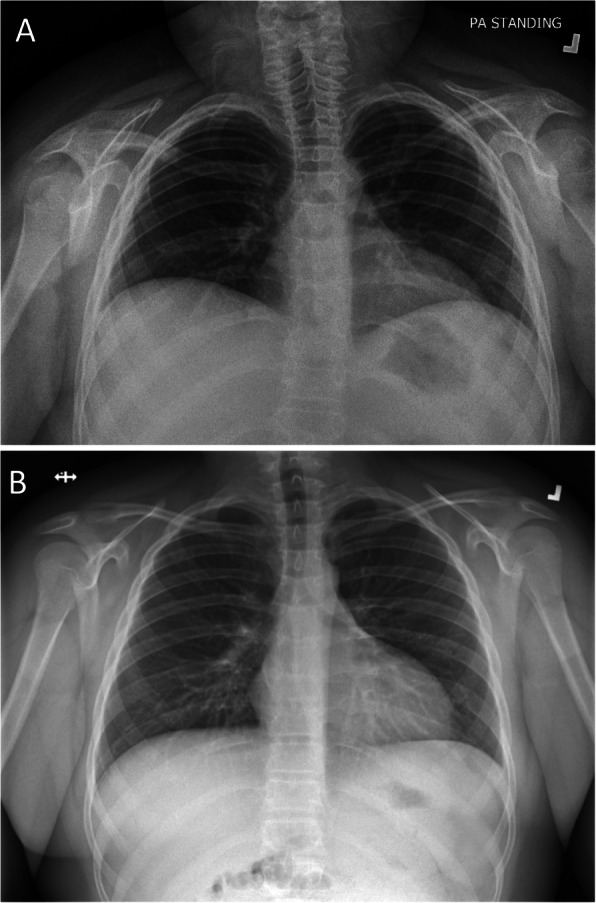
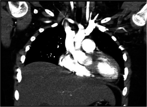
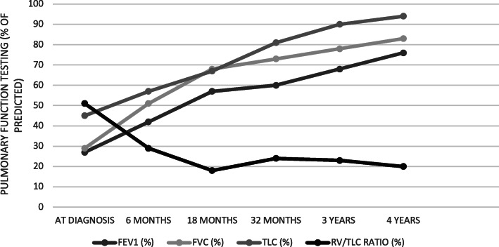
Case presentation: An 11-year-old boy presented with a malar rash, myositis, arthritis, oral ulcers, leukopenia, anemia, positive lupus autoantibodies and Class II nephritis. He was diagnosed with SLE and treated with corticosteroids, hydroxychloroquine, azathioprine, and subsequently mycophenolate with symptom resolution. At age 14, his SLE flared coincident with a viral chest infection. He presented with a malar rash, polyarthritis, increased proteinuria and pleuritis which all improved with corticosteroids and ongoing treatment with mycophenolate. Six weeks later he presented with severe dyspnea, markedly decreased lung volumes, but otherwise normal chest X-ray (CXR) and high-resolution chest computed tomography (HRCT). He was found to have severely restricted PFTs (FEV1 27%, FVC 29%; TLC 43%). After additional investigations including echocardiography, pulmonary CT angiography, and diaphragmatic fluoroscopy, he was diagnosed with SLS and treated with rituximab and methylprednisolone. At 1 month his symptoms had improved, but he still had dyspnea with exertion and severely restricted PFTs. At 6 months his FVC and TLC had improved to 51 and 57% respectively, and were 83 and 94% respectively at 4 years. He had returned to all baseline activities, including competitive hockey.
Conclusions: Although extremely rare, it is important to recognize SLS as a possible cause of dyspnea and chest pain in a child with SLE. Optimal treatment strategies are unknown. This is the second reported case of a child treated with rituximab for SLS who recovered normal lung function. International lupus registries should carefully document the occurrence, treatment and outcome of patients with SLS to help determine the optimal treatment for this rare complication.
Keywords: Child; Lung; Lupus; Rituximab; SLE.
Conflict of interest statement
The authors declare that they have no competing interests.
Figures
References
-
- Duron L, Cohen-Aubart F, Diot E, Borie R, Abad S, Richez C, et al. Shrinking lung syndrome associated with systemic lupus erythematosus: a multicenter collaborative study of 15 new cases and a review of the 155 cases in the literature focusing on treatment response and long-term outcomes. Autoimmun Rev. 2016;15:994–1000. doi: 10.1016/j.autrev.2016.07.021. - DOI - PubMed
Publication types
MeSH terms
Substances
LinkOut - more resources
Full Text Sources
Other Literature Sources
Medical
