

Combined treatment with enteric neural stem cells and chondroitinase ABC reduces spinal cord lesion pathology
- PMID: 33407795
- PMCID: PMC7789480
- DOI: 10.1186/s13287-020-02031-9
Combined treatment with enteric neural stem cells and chondroitinase ABC reduces spinal cord lesion pathology
Abstract
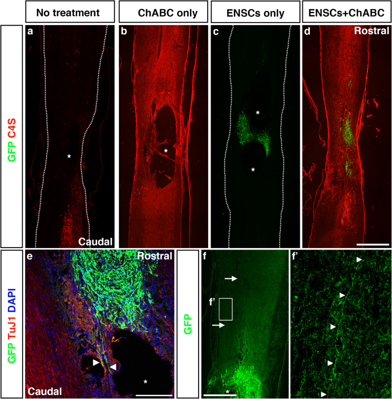
Background: Spinal cord injury (SCI) presents a significant challenge for the field of neurotherapeutics. Stem cells have shown promise in replenishing the cells lost to the injury process, but the release of axon growth-inhibitory molecules such as chondroitin sulfate proteoglycans (CSPGs) by activated cells within the injury site hinders the integration of transplanted cells. We hypothesised that simultaneous application of enteric neural stem cells (ENSCs) isolated from the gastrointestinal tract, with a lentivirus (LV) containing the enzyme chondroitinase ABC (ChABC), would enhance the regenerative potential of ENSCs after transplantation into the injured spinal cord.
Methods: ENSCs were harvested from the GI tract of p7 rats, expanded in vitro and characterised. Adult rats bearing a contusion injury were randomly assigned to one of four groups: no treatment, LV-ChABC injection only, ENSC transplantation only or ENSC transplantation+LV-ChABC injection. After 16 weeks, rats were sacrificed and the harvested spinal cords examined for evidence of repair.
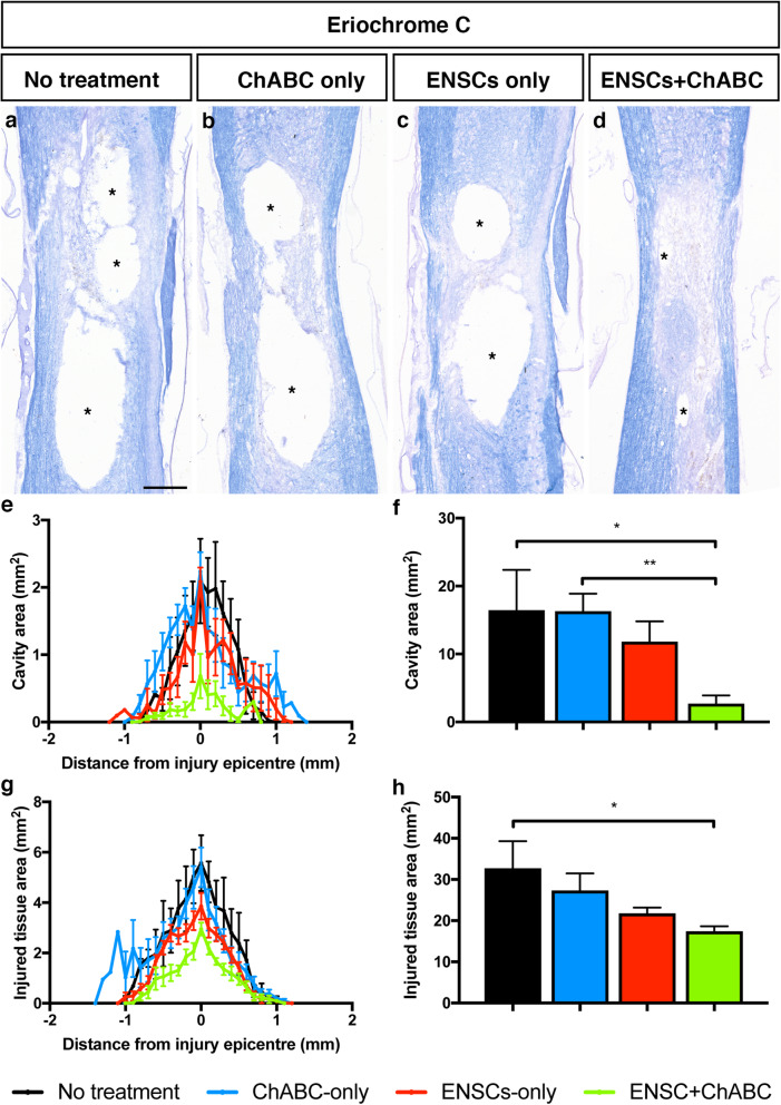
Results: ENSC cultures contained a variety of neuronal subtypes suitable for replenishing cells lost through SCI. Following injury, transplanted ENSC-derived cells survived and ChABC successfully degraded CSPGs. We observed significant reductions in the injured tissue and cavity area, with the greatest improvements seen in the combined treatment group. ENSC-derived cells extended projections across the injury site into both the rostral and caudal host spinal cord, and ENSC transplantation significantly increased the number of cells extending axons across the injury site. Furthermore, the combined treatment resulted in a modest, but significant functional improvement by week 16, and we found no evidence of the spread of transplanted cells to ectopic locations or formation of tumours.
Conclusions: Regenerative effects of a combined treatment with ENSCs and ChABC surpassed either treatment alone, highlighting the importance of further research into combinatorial therapies for SCI. Our work provides evidence that stem cells taken from the adult gastrointestinal tract, an easily accessible source for autologous transplantation, could be strongly considered for the repair of central nervous system disorders.
Keywords: ChABC; Enteric neural stem cells; Spinal cord injury; Stem cells.
Conflict of interest statement
The authors declare that they have no competing interests.
Figures


Similar articles
-
Neural stem cell mediated recovery is enhanced by Chondroitinase ABC pretreatment in chronic cervical spinal cord injury.PLoS One. 2017 Aug 3;12(8):e0182339. doi: 10.1371/journal.pone.0182339. eCollection 2017. PLoS One. 2017. PMID: 28771534 Free PMC article.
-
The combined application of human adipose derived stem cells and Chondroitinase ABC in treatment of a spinal cord injury model.Neuropeptides. 2017 Feb;61:39-47. doi: 10.1016/j.npep.2016.07.004. Epub 2016 Jul 28. Neuropeptides. 2017. PMID: 27484347
-
Effect of the combination of mesenchymal stromal cells and chondroitinase ABC on chronic spinal cord injury.Cytotherapy. 2015 Oct;17(10):1374-83. doi: 10.1016/j.jcyt.2015.05.012. Epub 2015 Jul 15. Cytotherapy. 2015. PMID: 26188966
-
Manipulating the glial scar: chondroitinase ABC as a therapy for spinal cord injury.Brain Res Bull. 2011 Mar 10;84(4-5):306-16. doi: 10.1016/j.brainresbull.2010.06.015. Epub 2010 Jul 8. Brain Res Bull. 2011. PMID: 20620201 Review.
-
The role of chondroitinase as an adjuvant to peripheral nerve repair.Cells Tissues Organs. 2014;200(1):59-68. doi: 10.1159/000369449. Epub 2015 Mar 4. Cells Tissues Organs. 2014. PMID: 25766067 Review.
Cited by
-
Recent Advances in the Treatment of Spinal Cord Injury.Arch Bone Jt Surg. 2024;12(6):380-399. doi: 10.22038/ABJS.2023.73944.3424. Arch Bone Jt Surg. 2024. PMID: 38919744 Free PMC article. Review.
-
Chondroitinase ABC combined with Schwann cell transplantation enhances restoration of neural connection and functional recovery following acute and chronic spinal cord injury.Neural Regen Res. 2025 May 1;20(5):1467-1482. doi: 10.4103/NRR.NRR-D-23-01338. Epub 2024 Jun 3. Neural Regen Res. 2025. PMID: 39075913 Free PMC article.
-
Bridging the gap of axonal regeneration in the central nervous system: A state of the art review on central axonal regeneration.Front Neurosci. 2022 Nov 9;16:1003145. doi: 10.3389/fnins.2022.1003145. eCollection 2022. Front Neurosci. 2022. PMID: 36440273 Free PMC article. Review.
-
The roles of neural stem cells in myelin regeneration and repair therapy after spinal cord injury.Stem Cell Res Ther. 2024 Jul 8;15(1):204. doi: 10.1186/s13287-024-03825-x. Stem Cell Res Ther. 2024. PMID: 38978125 Free PMC article. Review.
-
A Localized Materials-Based Strategy to Non-Virally Deliver Chondroitinase ABC mRNA Improves Hindlimb Function in a Rat Spinal Cord Injury Model.Adv Healthc Mater. 2022 Oct;11(19):e2200206. doi: 10.1002/adhm.202200206. Epub 2022 Aug 25. Adv Healthc Mater. 2022. PMID: 35882512 Free PMC article.
References
Publication types
MeSH terms
Substances
Grants and funding
LinkOut - more resources
Full Text Sources
Other Literature Sources
Medical
