. 2021 Jan 6;13(1):1.
doi: 10.1186/s13148-020-00979-2.

Micro-RNA-125a mediates the effects of hypomethylating agents in chronic myelomonocytic leukemia

Affiliations

Micro-RNA-125a mediates the effects of hypomethylating agents in chronic myelomonocytic leukemia

Johannes Lorenz Berg et al. Clin Epigenetics. .

Abstract

Background: Chronic myelomonocytic leukemia (CMML) is an aggressive hematopoietic malignancy that arises from hematopoietic stem and progenitor cells (HSPCs). Patients with CMML are frequently treated with epigenetic therapeutic approaches, in particular the hypomethylating agents (HMAs), azacitidine (Aza) and decitabine (Dec). Although HMAs are believed to mediate their efficacy via re-expression of hypermethylated tumor suppressors, knowledge about relevant HMA targets is scarce. As silencing of tumor-suppressive micro-RNAs (miRs) by promoter hypermethylation is a crucial step in malignant transformation, we asked for a role of miRs in HMA efficacy in CMML.

Results: Initially, we performed genome-wide miR-expression profiling in a KrasG12D-induced CMML mouse model. Selected candidates with prominently decreased expression were validated by qPCR in CMML mice and human CMML patients. These experiments revealed the consistent decrease in miR-125a, a miR with previously described tumor-suppressive function in myeloid neoplasias. Furthermore, we show that miR-125a downregulation is caused by hypermethylation of its upstream region and can be reversed by HMA treatment. By employing both lentiviral and CRISPR/Cas9-based miR-125a modification, we demonstrate that HMA-induced miR-125a upregulation indeed contributes to mediating the anti-leukemic effects of these drugs. These data were validated in a clinical context, as miR-125a expression increased after HMA treatment in CMML patients, a phenomenon that was particularly pronounced in cases showing clinical response to these drugs.

Conclusions: Taken together, we report decreased expression of miR-125a in CMML and delineate its relevance as mediator of HMA efficacy within this neoplasia.

Keywords: Azacitidine; Chronic myelomonocytic leukemia; Hypomethylating agent; Tumor suppressor; miRNA.


Conflict of interest statement

The authors declare that they have no competing interests.

Figures

Fig. 1
Fig. 1
Deregulation of miR expression levels in a murine model of CMML. a To screen for aberrant miR expression profiles in CMML, we performed miR-microarray analysis in CD-11b/Ly-6G/c-Kit+ HSPCs isolated from the bone marrow of mice with a Ras-induced CMML-like MPD. Therefore, four Mx1-Cre+/KrasG12D mice and four KrasWt control mice were injected with pIpC as outlined in the materials and methods section. Mice were killed and analyzed 6 weeks later. The development of the CMML-like MPD at this time was verified as outlined in Additional file 1: Fig S13. b Volcano plot showing deregulated miRs in CMML-HSPCs of Mx1-Cre+/KrasG12D-mutated mice. The horizontal axis depicts the x-fold expression change in Mx1-Cre+/KrasG12D-mutated animals and transformed P-values are depicted on the vertical axis. miRs with higher expression in KrasG12D-mutated HSPCs are displayed on the right, whereas miRs with decreased expression levels in KrasG12D-mutated HSPCs are displayed on the left. cd qPCR validation of the miR microarray results shows decreased miR-125a, miR-150, and miR-26a expression in CD-11b/Ly-6G/c-Kit+ (c, n = 3) and Lin/Sca-1+/c-Kit+ (d, n = 4) HSPC compartments of Mx1-Cre+/KrasG12D mice. At least three mice per group were analyzed. The dot plots show the relative miR expression with the median indicated as horizontal line. One KrasWt mouse was always used as a calibrator and its expression is set to 1. Group differences were assessed by t test. Wt, wildtype;
Fig. 2
Fig. 2
miR-125a expression is decreased in primary CMML patient specimens. a The box plots show the relative miR-125a, miR-150, and miR-26a expression levels in 36 CMML patient specimens compared to six CD34+ HSPCs. b The relative expression level of miR-125a in 36 CMML patients compared to six whole BM specimens of healthy donors and patients with lymphatic diseases without BM affection is displayed. The graphs denote the miR expression normalized to a calibrator, set to 1 (U937 for miR-125a and miR-26a; GDM-1 for miR-150). Differences between the groups were assessed with the Mann–Whitney U test. CMML, chronic myelomonocytic leukemia; BM, bone marrow; HSPCs, hematopoietic stem and progenitor cells
Fig. 3
Fig. 3
Decreased miR-125a expression is caused by hypermethylation of its upstream/promoter region and can be increased by HMA treatment. a miR-125a expression was assessed by qPCR in the myeloid cell lines THP1, U937, and NB4 after treatment with 5 µM decitabine for 48 h. For comparison of the different conditions, respective control situations (treated with the empty dissolvent only) were set at a value of 1. The relative increase in miR-125a expression in the decitabine-treated conditions was calculated as the ratio of decitabine-treated to control-treated expression levels. b miR-125a expression after treatment with 2.5 µM azacitidine for 24 h. The relative increase in miR-125a expression in the azacitidine-treated conditions compared to controls is displayed. Graphs denote the mean ± SD of at least three independent experiments. Comparisons against the control condition were performed using a one-sample t test against a reference value of 1. Of note, the increase in miR-125a expression could also be observed after incubation with a lower azacitidine concentration (1 µM; Additional file 1: Fig. S4) c Eight CpG-sites within a CpG-rich region upstream of miR-125a [23] were studied by bisulfite sequencing in U937 cells before and after treatment with 2.5 µM azacitidine for 24 h. This region roughly correlates to the miR-125a promoter region [52]. The panel on the left demonstrates the percentage of methylated sequencing reads for each CpG site studied; the panel on the right depicts the mean percentage of methylation across all eight CpG sites ± SD. Statistical differences were assessed with the t test. Aza, azacitidine; Dec, decitabine
Fig. 4
Fig. 4
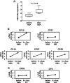
miR-125a expression increases in CMML patients after the therapy with HMAs. a Box plot depicting the miR-125a expression levels in seven paired CMML patient specimens collected before and after treatment with HMAs. miR-125a expression was analyzed by qPCR and displayed as the log-transformed x-fold expression of the calibrator (U937 cells). Differences between expression values before and after treatment were assessed with the paired Wilcoxon signed-rank test. b,c, miR-125a expression levels in individual CMML patients with (b) or without (c) clinical response to HMA therapy. miR-125a expression levels assessed before HMA treatment were used as controls and set to 1. miR-125a levels after HMA treatment are displayed as x-fold change to the respective control sample. HMA hypomethylating agent; CP, CMML patient, Dec, decitabine; Aza, azacitidine, CR, complete response; MR, marrow response; CB, clinical benefit; PR, partial response; PD, progressive disease; NR, no response
Fig. 5
Fig. 5
The anti-leukemic effects of HMAs are partly mediated by increasing the expression of miR-125a. a THP1 cells were transiently transfected with a miR-125a-specific hairpin inhibitor (THP1 miR-125a KD) and scrambled control (THP1 control KD), respectively. b miR-125a was deleted in U937 by employing the CRISPR/Cas9 technology (U937 miR-125a KO); parental U937 cells were used as controls. Apoptosis was measured after 24-h treatment with Aza (2.5 µM for THP1, 5 µM for U937; indicated as +) or empty dissolvent (indicated as −) by Annexin-V/7AAD assays. The respective control situations (treated with the empty dissolvent only) were set at a value of 1, and the relative increase in apoptosis in the Aza-treated conditions was calculated as the ratio of Aza-treated to control-treated cells. The graphs represent the mean ± SD of at least three independent experiments. Statistical significance was evaluated using a paired Student’s t test

References

    1. Arber DA, Orazi A, Hasserjian R, Thiele J, Borowitz MJ, Le Beau MM, et al. The 2016 revision to the World Health Organization classification of myeloid neoplasms and acute leukemia. Blood. 2016;127:2391–2405. - PubMed
    1. Itzykson R, Fenaux P, Bowen D, Cross NCP, Cortes J, De Witte T, et al. Diagnosis and treatment of chronic myelomonocytic leukemias in adults: recommendations from the European Hematology Association and the European LeukemiaNet. Hemasphere. 2018;2:e150. doi: 10.1097/HS9.0000000000000150. - DOI - PMC - PubMed
    1. Patnaik MM, Tefferi A. Chronic Myelomonocytic leukemia: 2020 update on diagnosis, risk-stratification and management. Am J Hematol. 2020;95:97–115. doi: 10.1002/ajh.25684. - DOI - PubMed
    1. Geissler K, Jager E, Barna A, Gurbisz M, Marschon R, Graf T, et al. The Austrian biodatabase for chronic myelomonocytic leukemia (ABCMML): a representative and useful real-life data source for further biomedical research. Wien Klin Wochenschr. 2019;131:410–418. doi: 10.1007/s00508-019-1526-1. - DOI - PMC - PubMed
    1. Diesch J, Zwick A, Garz AK, Palau A, Buschbeck M, Gotze KS. A clinical-molecular update on azanucleoside-based therapy for the treatment of hematologic cancers. Clin Epigenetics. 2016;8:71016-0237-y. eCollection 2016. - PMC - PubMed

Publication types

MeSH terms