. 2021 Jan 6;12(1):4.
doi: 10.1186/s13287-020-02072-0.

Hypoxic hUCMSC-derived extracellular vesicles attenuate allergic airway inflammation and airway remodeling in chronic asthma mice

Affiliations

Hypoxic hUCMSC-derived extracellular vesicles attenuate allergic airway inflammation and airway remodeling in chronic asthma mice

Liyang Dong et al. Stem Cell Res Ther. .

Abstract

Background: As one of the main functional forms of mesenchymal stem cells (MSCs), MSC-derived extracellular vesicles (MSC-EVs) have shown an alternative therapeutic option in experimental models of allergic asthma. Oxygen concentration plays an important role in the self-renewal, proliferation, and EV release of MSCs and a recent study found that the anti-asthma effect of MSCs was enhanced by culture in hypoxic conditions. However, the potential of hypoxic MSC-derived EVs (Hypo-EVs) in asthma is still unknown.

Methods: BALB/c female mice were sensitized and challenged with ovalbumin (OVA), and each group received PBS, normoxic human umbilical cord MSC-EVs (Nor-EVs), or Hypo-EVs weekly. After treatment, the animals were euthanized, and their lungs and bronchoalveolar lavage fluid (BALF) were collected. With the use of hematoxylin and eosin (HE), periodic acid-Schiff (PAS) and Masson's trichrome staining, enzyme-linked immune sorbent assay (ELISA), Western blot analysis, and real-time PCR, the inflammation and collagen fiber content of airways and lung parenchyma were investigated.

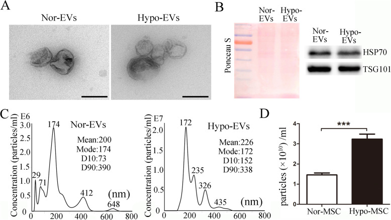
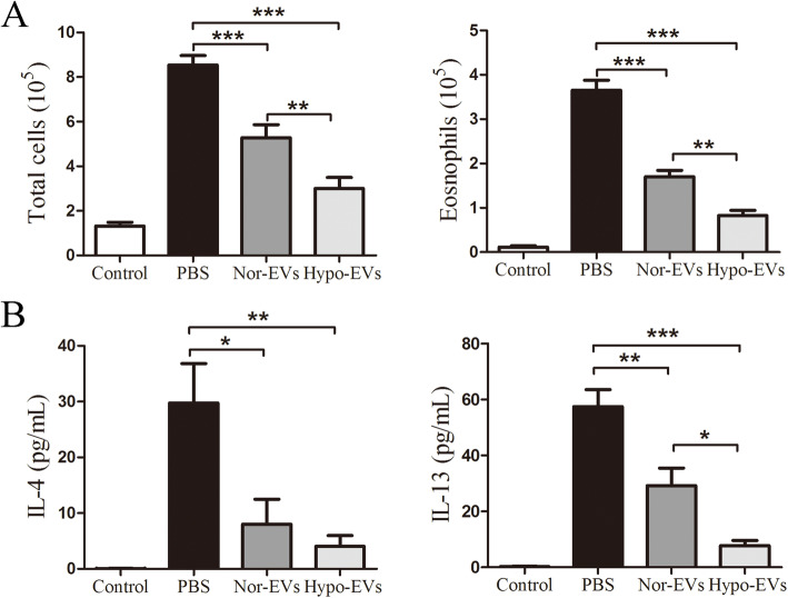
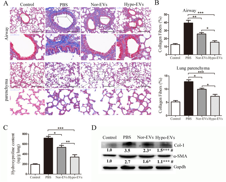
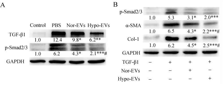
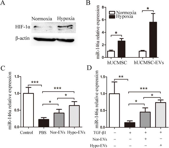
Results: Hypoxic environment can promote human umbilical cord MSCs (hUCMSCs) to release more EVs. In OVA animals, the administration of Nor-EVs or Hypo-EVs significantly ameliorated the BALF total cells, eosinophils, and pro-inflammatory mediators (IL-4 and IL-13) in asthmatic mice. Moreover, Hypo-EVs were generally more potent than Nor-EVs in suppressing airway inflammation in asthmatic mice. Compared with Nor-EVs, Hypo-EVs further prevented mouse chronic allergic airway remodeling, concomitant with the decreased expression of pro-fibrogenic markers α-smooth muscle actin (α-SMA), collagen-1, and TGF-β1-p-smad2/3 signaling pathway. In vitro, Hypo-EVs decreased the expression of p-smad2/3, α-SMA, and collagen-1 in HLF-1 cells (human lung fibroblasts) stimulated by TGF-β1. In addition, we showed that miR-146a-5p was enriched in Hypo-EVs compared with that in Nor-EVs, and Hypo-EV administration unregulated the miR-146a-5p expression both in asthma mice lung tissues and in TGF-β1-treated HLF-1. More importantly, decreased miR-146a-5p expression in Hypo-EVs impaired Hypo-EV-mediated lung protection in OVA mice.

Conclusion: Our findings provided the first evidence that hypoxic hUCMSC-derived EVs attenuated allergic airway inflammation and airway remodeling in chronic asthma mice, potentially creating new avenues for the treatment of asthma.

Keywords: Asthma; Extracellular vesicles; Human umbilical cord mesenchymal stem cells; Hypoxia; Lung injury.


Conflict of interest statement

There are no conflicts of interest.

Figures

Fig. 1
Fig. 1
Characterization of Nor-EVs and Hypo-EVs. a Transmission electron micrographs of Nor-EVs and Hypo-EVs. Scale bar = 200 nm. b Western blot analysis of TSG101 and HSP60 expression in Nor-EVs and Hypo-EVs (n = 3). Ponceau S staining served as a loading control. c NTA analysis of Nor-EVs and Hypo-EVs revealed that EVs from the two groups exhibit similar size ranges (n = 3). d NTA counted the production of EVs derived from normoxia-cultured hUCMSCs (Nor-MSC) and EVs from hypoxia-treated hUCMSCs (Hypo-MSC) (n = 3). ***P < 0.001
Fig. 2
Fig. 2
Administration of Hypo-EVs attenuated OVA-induced chronic airway inflammation in mice. a Schematic illustration of the establishment of mouse chronic allergic airway inflammation model. b Distribution of DIR-labeled EVs in OVA-induced mice lung after tail vein administration (26 day injection and 28 day detected). Representative ex vivo bioluminescence images of mice lungs from different groups (left), and the quantification of the relative fluorescence intensity in the lungs (right; n = 3 from three independent experiments). c Representative photographs of HE- and PAS-stained lung sections from each group (black bar = 200 μm). d, e The inflammatory infiltration and goblet cell hyperplasia were quantified by HE and PAS scores (n = 6). *P < 0.05, **P < 0.01, ***P < 0.001
Fig. 3
Fig. 3
Hypo-EV treatment reduced inflammatory cell infiltration and inflammatory cytokines in the BALF. a Statistical analysis of the total inflammatory cells (left) and eosinophils (right) in the BALF (n = 6). b Statistical analysis of IL-4 (left) and IL-13 (right) levels in the BALF as measured by ELISA (n = 6). *P < 0.05, **P < 0.01, ***P < 0.001
Fig. 4
Fig. 4
Hypo-EV treatment prevented the airway and lung parenchyma fibrosis in chronic asthma mice. a Representative photomicrographs of airway (upper panels) and lung parenchyma (lower panels) stained with Masson trichrome staining (black bar = 200 μm). b Percentage of collagen fiber content in airway and lung parenchyma (n = 6). *P < 0.05, **P < 0.01, ***P < 0.001. c Collagen levels in lung tissue determined by hydroxyproline assay (n = 6). *P < 0.05, **P < 0.01, ***P < 0.001. d Representative Western blots of Col-1 and α-SMA (GAPDH was used as protein loading control) in the lungs from each group (n = 4), and band intensities were normalized against the corresponding GAPDH and the numbers are presented as fold increase over the control group. *P < 0.05, ***P < 0.01 vs. PBS group; #P < 0.05 vs. Nor-EV group
Fig. 5
Fig. 5
Effect of Hypo-EVs on the expression of TGF-β1-phosphorylated (p)-Smad2/3 in lung tissues and in TGF-β1-treated HLF-1 cells. a Representative Western blots of TGF-β1 and p-Smad2/3 (GAPDH was used as protein loading control) in the lungs from each group (n = 4), and band intensities were normalized against the corresponding GAPDH and the numbers are presented as fold increase over the control group. *P < 0.05, **P < 0.01, ***P < 0.001 vs. PBS group; #P < 0.05 vs. Nor-EV group. b The expression of p-Smad2/3, α-SMA, collogen-1, and GAPDH proteins in TGF-β1-treated HLF-1 cells were determined by Western blotting (n = 3 from three independent experiments). Band intensities were normalized against the corresponding GAPDH (protein loading control) and the numbers are presented as fold increase over the control group. *P < 0.05, ***P < 0.001 vs. TGF-β1 group; #P < 0.05 vs. TGF-β1+Nor-EV group
Fig. 6
Fig. 6
The expression of miR-146a-5p in Hypo-EVs. a The protein expression of HIF-1α in hUCMSCs under normoxic and hypoxic conditions. b The expression of miR-146a-5p in hUCMSCs or EVs were detected by real-time PCR (n = 3). c MiR-146a-5p expression in the lung tissues of the control, PBS, Nor-EV, and Hypo-EV group (n = 6). d The expression of miR-146a-5p in HLF-1 cells that were treated with Nor-EVs or Hypo-EVs were detected by real-time PCR (n = 3). *P < 0.05, **P < 0.01, ***P < 0.001
Fig. 7
Fig. 7
Inhibition of miR-146a-5p in Hypo-EVs impaired Hypo-EV-mediated lung protection in a mice model of asthma. a miR-146a-5p expression was detected by real-time PCR in EVs isolated from the hypoxic medium of hUCMSCs treated with 100 nM inhibitor-146a-5p or negative control (NC) (n = 3). b Expression of miR-145a-5p in lung tissue after injection of miR146aKD-Hypo-EVs or NC-Hypo-EVs into the asthma mice (n = 6). c Representative photographs of HE- and PAS-stained lung sections from each group (black bar = 200 μm). d The inflammatory infiltration and goblet cell hyperplasia were quantified by HE and PAS scores (n = 6). e Statistical analysis of the total inflammatory cells and eosinophils in the BALF (n = 6). f Statistical analysis of IL-4 and IL-13 levels in the BALF as measured by ELISA (n = 6). g Representative photomicrographs of airway and parenchyma stained with Masson trichrome staining (black bar = 200 μm). h Percentage of collagen fiber content in airway and lung parenchyma (n = 6). i Representative Western blots of TGF-β1, p-Smad2/3, α-SMA, and collogen-1 in the lungs from each group (n = 3), and band intensities were normalized against the corresponding GAPDH and the numbers are presented as fold increase over the NC-Hypo-EV group. j The expression of p-Smad2/3, α-SMA, and collogen-1 in TGF-β1-treated HLF-1 cells were determined by Western blotting (n = 3). Band intensities were normalized against the corresponding GAPDH and the numbers are presented as fold increase over the NC-Hypo-EV group. *P < 0.05, **P < 0.01, ***P < 0.001

Similar articles

Cited by

References

    1. Backman H, Raisanen P, Hedman L, et al. Increased prevalence of allergic asthma from 1996 to 2006 and further to 2016-results from three population surveys. Clin Exp Allergy. 2017;47(11):1426–1435. doi: 10.1111/cea.12963. - DOI - PubMed
    1. Lambrecht BN, Hammad H. The immunology of asthma. Nat Immunol. 2015;16:45–56. doi: 10.1038/ni.3049. - DOI - PubMed
    1. Papierniak ES, Lowenthal DT, Harman E. Novel therapies in asthma: leukotriene antagonists, biologic agents, and beyond. Am J Ther. 2013;20:79–103. doi: 10.1097/MJT.0b013e31826915c2. - DOI - PubMed
    1. Zhang LB, He M. Effect of mesenchymal stromal (stem) cell (MSC) transplantation in asthmatic animal models: a systematic review and meta-analysis. Pulm Pharmacol Ther. 2019;54:39–52. doi: 10.1016/j.pupt.2018.11.007. - DOI - PubMed
    1. Harrell CR, Sadikot R, Pascual J, et al. Mesenchymal stem cell-based therapy of inflammatory lung diseases: current understanding and future perspectives. Stem Cells Int. 2019;2019:4236973. - PMC - PubMed

Publication types