. 2021 Feb 24;95(6):e02369-20.
doi: 10.1128/JVI.02369-20. Print 2021 Feb 24.

Phosphorylation of Influenza A Virus NS1 at Serine 205 Mediates Its Viral Polymerase-Enhancing Function

Affiliations

Phosphorylation of Influenza A Virus NS1 at Serine 205 Mediates Its Viral Polymerase-Enhancing Function

Amol Patil et al. J Virol. .

Abstract

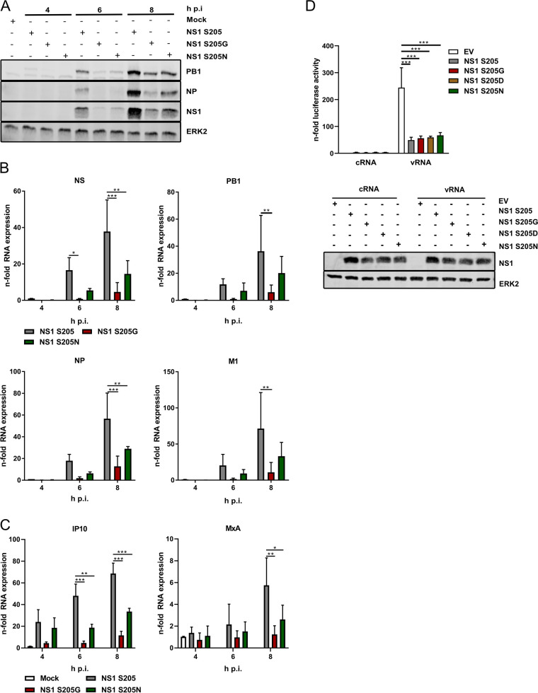
Influenza A virus (IAV) nonstructural protein 1 (NS1) is a protein with multiple functions that are regulated by phosphorylation. Phosphoproteomic screening of H1N1 virus-infected cells revealed that NS1 was phosphorylated at serine 205 in intermediate stages of the viral life cycle. Interestingly, S205 is one of six amino acid changes in NS1 of post-pandemic H1N1 viruses currently circulating in humans compared to the original swine-origin 2009 pandemic (H1N1pdm09) virus, suggesting a role in host adaptation. To identify NS1 functions regulated by S205 phosphorylation, we generated recombinant PR8 H1N1 NS1 mutants with S205G (nonphosphorylatable) or S205N (H1N1pdm09 signature), as well as H1N1pdm09 viruses harboring the reverse mutation NS1 N205S or N205D (phosphomimetic). Replication of PR8 NS1 mutants was attenuated relative to wild-type (WT) virus replication in a porcine cell line. However, PR8 NS1 S205N showed remarkably higher attenuation than PR8 NS1 S205G in a human cell line, highlighting a potential host-independent advantage of phosphorylatable S205, while an asparagine at this position led to a potential host-specific attenuation. Interestingly, PR8 NS1 S205G did not show polymerase activity-enhancing functions, in contrast to the WT, which can be attributed to diminished interaction with cellular restriction factor DDX21. Analysis of the respective kinase mediating S205 phosphorylation indicated an involvement of casein kinase 2 (CK2). CK2 inhibition significantly reduced the replication of WT viruses and decreased NS1-DDX21 interaction, as observed for NS1 S205G. In summary, NS1 S205 is required for efficient NS1-DDX21 binding, resulting in enhanced viral polymerase activity, which is likely to be regulated by transient phosphorylation.IMPORTANCE Influenza A viruses (IAVs) still pose a major threat to human health worldwide. As a zoonotic virus, IAV can spontaneously overcome species barriers and even reside in new hosts after efficient adaptation. Investigation of the functions of specific adaptational mutations can lead to a deeper understanding of viral replication in specific hosts and can probably help to find new targets for antiviral intervention. In the present study, we analyzed the role of NS1 S205, a phosphorylation site that was reacquired during the circulation of pandemic H1N1pdm09 "swine flu" in the human host. We found that phosphorylation of human H1N1 virus NS1 S205 is mediated by the cellular kinase CK2 and is needed for efficient interaction with human host restriction factor DDX21, mediating NS1-induced enhancement of viral polymerase activity. Therefore, targeting CK2 activity might be an efficient strategy for limiting the replication of IAVs circulating in the human population.

Keywords: NS1; influenza virus; protein phosphorylation; viral polymerase activity.


Figures

FIG 1
FIG 1
PR8 NS1 S205 is phosphorylated in viral replication. (A, B) Amino acid prevalence at NS1 position 205 in all human H1N1 viruses (A) and swine H1N1 isolates (B) from Northern temperate regions from 1930 until 2020 (n, 6 to 5,317 isolates/time frame). (C) Prevalence of a serine at amino acid position 205 in the viral NS1 protein. Human H3N2 and H2N2 isolates, swine H1N2 and H3N2 isolates, and avian isolates of different subtypes isolated from 1902 to 2020 were analyzed (n, 125 to 22,092). (D) Relative phosphorylation of the tryptic peptide of NS1 showing the amount of phosphorylated S205 normalized to total NS1 protein amounts. (E) A549 cells were transfected with WT PR8 NS1 or the S205G substitution mutant. At 24 h p.t., cells were infected with WT PR8 (MOI, 5) for 6 or 8 h. (Left) Phosphoserine-containing proteins were immunoprecipitated (IP) by using pS-specific antibodies, and the precipitation of NS1 was determined by Western blot analysis. Blots are representative of the results of three independent experiments. The phosphorylation levels of NS1 were analyzed by densitometry and normalized to the respective NS1 amounts. (Right) Shown are the levels of phosphorylated NS1 (+ standard deviations) as percentages of the phosphorylation level of NS1 S205 at 6 h p.i. Statistical significance was analyzed by one-way analysis of variance followed by Dunn’s multiple-comparison test.
FIG 2
FIG 2
A serine at position 205 of the NS1 protein facilitates efficient viral replication. (A to E) Human A549 cells (A, C), porcine WSL-R-HP cells (B, D), or HBEpCs (E) were infected with WT PR8, PR8 NS1 S205G, or PR8 NS1 S205N (A, B) or with WT H1N1pdm09, H1N1pdm09 NS1 N205S, or H1N1pdm09 NS1 N205D (C, D, E) at an MOI of 0.1, and viral replication was analyzed at the indicated time points (A to D) or 24 h p.i. (E). Amounts of viral progeny were analyzed by standard plaque assays and are presented as means ± standard deviations of results from three independent experiments (A) or as results from one experiment representative of two independent experiments (B, C, D). Virus titers obtained after infection of HBEpCs are expressed as mean percentages of the WT H1N1pdm09 NS1 titer ± standard deviations from three independent experiments (E). Statistical significance was analyzed by one-way (E) or two-way (A to D) analysis of variance followed by Tukey’s multiple-comparison test (***, P ≤ 0.001).
FIG 3
FIG 3
NS1 S205 substitution mutants show decreased viral protein expression that is independent of the IFN-antagonistic function of NS1. (A, B, C) A549 cells were infected with WT PR8 or substitution mutants at an MOI of 5 and were analyzed 4, 6, and 8 h p.i. for the expression of viral proteins (A) or viral mRNA and cRNA (B) as well as cellular innate immune responses (C). (A) Expression of viral proteins PB1, NP, and NS1 was determined by Western blotting. ERK2 served as a loading control. Blots are representative of the results of three independent experiments. (B, C) The expression of different viral and cellular mRNAs was analyzed by qRT-PCR and is presented as the mean n-fold induction + standard deviation for three independent experiments. Values were normalized to those for WT virus infection at 4 h p.i. (B) or mock-infected controls (C). Statistical analysis was performed by two-way analysis of variance followed by Tukey’s multiple-comparison test (*, P ≤ 0.05; **, P ≤ 0.01; ***, P ≤ 0.001). (D) Impact of PR8 NS1 S205 substitution on IFN-β promoter activity. Vero cells were transfected with a luciferase reporter construct expressed under the control of the IFN-β promoter in combination with an empty vector (EV), PR8 NS1 S205, or substitution mutants for 24 h. Cells were stimulated with 500 ng of total RNA isolated from infected A549 cells (8 h; MOI, 5; vRNA). Total RNA from uninfected A549 cells was used as a control (cRNA). At 5 h poststimulation, promoter activity was measured by a luciferase assay, and the results are presented as the mean n-fold activity (+ standard deviation) from three independent experiments. Values were normalized to those for the EV cRNA-stimulated controls. Statistical significance was analyzed by two-way analysis of variance followed by Sidak’s multiple-comparison test (***, P ≤ 0.001).
FIG 4
FIG 4
NS1-mediated enhancement of viral polymerase activity is orchestrated by interaction with cellular DDX21. (A, C) Effects of WT PR8 (A) or H1N1pdm09 (C) NS1 and substitution mutants on viral polymerase activity. Vero cells were transfected with plasmids encoding WT NS1 or different NS1 205 substitution mutants, PB1, PB2, PA, and NP, together with a plasmid that directs the expression of the firefly luciferase reporter RNA minigenome. At 24 h p.t., cells were lysed and firefly luciferase activity was measured. The results are presented as the mean relative polymerase activity (+ standard deviation) for three independent experiments normalized to NS1 expression levels and activity in empty-vector-expressing cells. Confirmation of efficient NS1 overexpression was performed by Western blot analysis. ERK2 served as a loading control for detection. Statistical significance was analyzed by one-way analysis of variance followed by Dunnett’s multiple-comparison test (*, P ≤ 0.05; **, P ≤ 0.01; ***, P ≤ 0.001). (B) A549 cells were infected at an MOI of 20 with WT PR8 NS1 or the PR8 NS1 S205G or S205N substitution mutant for 8 h. Cells were fixed, and the localization of viral proteins NS1 (green) and NP (red) was analyzed by indirect immunofluorescence. DAPI was used to stain the nuclei. Images are representative of three independent experiments. (D) Analysis of NS1–DDX21 interaction. Vero cells were transfected with Flag-tagged DDX21 in combination with Strep-tagged WT NS1 or S205 substitution mutants. At 24 h p.t., NS1 was pulled down by using Strep-Tactin beads. (Top) Coprecipitation of DDX21 was determined by using Flag-specific antibodies in Western blot analysis. (Bottom) Amounts of coprecipitated DDX21 were analyzed by densitometry of blots from three to six independent experiments and are presented as the level of interaction compared to that with WT NS1 (+ standard deviation). Statistical significance was analyzed by one-way analysis of variance followed by Dunn’s multiple-comparison test (*, P ≤ 0.05). (E) A549 cells were transfected with different DDX21-specific siRNAs or scrambled control siRNAs. At 48 h p.t., cells were infected at an MOI of 0.1 with WT PR8 NS1 or PR8 expressing NS1 S205G or NS1 S205N. (Top) Virus titers were determined 24 h p.i. by standard plaque assays and are presented as the mean virus titer (+ standard deviation) (left) or as the mean percentage, from three independent experiments, of the virus titer for WT infection of control siRNA-transfected cells (right). Statistical significance was analyzed by two-way analysis of variance followed by Tukey’s multiple-comparison test (*, P ≤ 0.05; ***, P ≤ 0.001). (Bottom) Efficient DDX21 knockdown was confirmed by Western blot analysis. ERK1/2 was used as a loading control, and NS1 detection served as a control for infection.
FIG 5
FIG 5
Cellular casein kinase 2 is required for efficient replication by catalyzing NS1 S205 phosphorylation, which is needed for DDX21 interaction. (A) Schematic representation of the CK2 recognition site in comparison to the sequence containing PR8 NS1 S205. (B) A549 cells were pretreated with the CK2-specific inhibitor DMAT (5 μM) or DMSO for 1 h. Cells were infected at an MOI of 0.1 with WT PR8 NS1 or PR8 expressing NS1 S205G and were treated with DMSO or DMAT (5 μM) for 24 h. Virus-containing supernatants were harvested, and viral titers were determined by standard plaque assays and are presented as means (+ standard deviations) from three independent experiments (left) or as the percentage of reduction in viral replication in DMAT-treated cells (right). The level of viral replication in DMSO-treated cells was arbitrarily set to 100%. Statistical significance was determined by using two-way analysis of variance followed by Sidak’s multiple-comparison test (*, P ≤ 0.05; **, P ≤ 0.01). (C) A549 cells were transfected with NS1 S205 or substitution mutants for 24 h. Subsequently, cells were preincubated with DMSO or DMAT (5 μM) for 1 h and were then infected with PR8 ΔNS1 (MOI = 1) in the presence of DMSO or DMAT. Expression levels of NP, M1, and PB1 mRNAs and cRNAs were analyzed 4 and 8 h p.i. by qRT-PCR and are presented as mean n-fold induction (+ standard deviation) from one experiment representative of two independent experiments. Values were normalized to those for cells overexpressing NS1 S205 at 4 h p.i. Statistical significance was analyzed by two-way analysis of variance followed by Tukey’s multiple-comparison test (**, P ≤ 0.01; ***, P ≤ 0.001). (D) Effects of CK2 inhibition on the NS1–DDX21 interaction. HEK293 cells were transfected with Flag-tagged DDX21 in combination with Strep-tagged WT NS1 or substitution mutants. At 18 h p.t., cells were treated with the CK2 inhibitor DMAT (5 μM) or DMSO for 6 h, and NS1 was pulled down by using Strep-Tactin Sepharose beads. Coprecipitation of DDX21 was determined by using Flag-specific antibodies, and efficient Strep-NS1 pulldown was confirmed by using NS1-specific antibodies in Western blot analysis. Blots are representative of the results of two independent experiments.

References

    1. Nogales A, Martinez-Sobrido L, Topham DJ, DeDiego ML. 2018. Modulation of innate immune responses by the influenza A NS1 and PA-X proteins. Viruses 10:708. doi:10.3390/v10120708. - DOI - PMC - PubMed
    1. Marc D. 2014. Influenza virus non-structural protein NS1: interferon antagonism and beyond. J Gen Virol 95:2594–2611. doi:10.1099/vir.0.069542-0. - DOI - PubMed
    1. Hale BG, Randall RE, Ortin J, Jackson D. 2008. The multifunctional NS1 protein of influenza A viruses. J Gen Virol 89:2359–2376. doi:10.1099/vir.0.2008/004606-0. - DOI - PubMed
    1. Mok BW, Song W, Wang P, Tai H, Chen Y, Zheng M, Wen X, Lau SY, Wu WL, Matsumoto K, Yuen KY, Chen H. 2012. The NS1 protein of influenza A virus interacts with cellular processing bodies and stress granules through RNA-associated protein 55 (RAP55) during virus infection. J Virol 86:12695–12707. doi:10.1128/JVI.00647-12. - DOI - PMC - PubMed
    1. Chen G, Liu CH, Zhou L, Krug RM. 2014. Cellular DDX21 RNA helicase inhibits influenza A virus replication but is counteracted by the viral NS1 protein. Cell Host Microbe 15:484–493. doi:10.1016/j.chom.2014.03.002. - DOI - PMC - PubMed

Publication types

MeSH terms

LinkOut - more resources