. 2020 Dec 21:12:559603.
doi: 10.3389/fnagi.2020.559603. eCollection 2020.

Dysfunction of the Glymphatic System Might Be Related to Iron Deposition in the Normal Aging Brain

Affiliations

Dysfunction of the Glymphatic System Might Be Related to Iron Deposition in the Normal Aging Brain

Wei Zhou et al. Front Aging Neurosci. .

Abstract

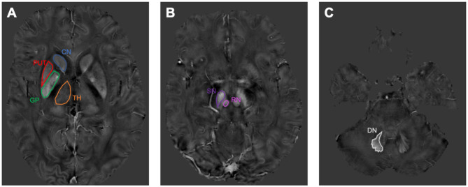
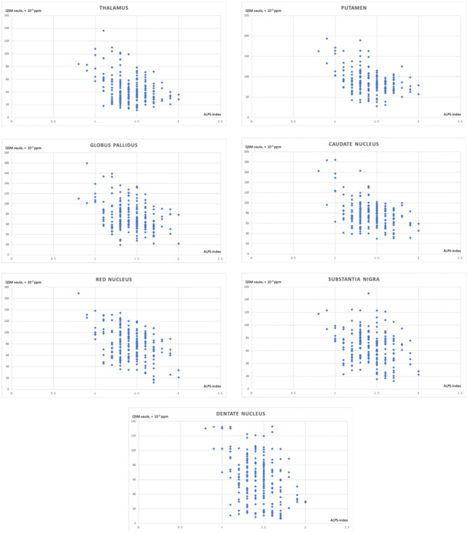
Objective: The study aims to detect the potential relationship between iron deposition and the function of the glymphatic system in the normal aging brain. Methods: We recruited 213 healthy participants. We evaluated the function of the glymphatic system using the index for diffusivity along the perivascular space (ALPS-index), assessed iron deposition on quantitative susceptibility mapping (QSM), and analyzed their relationship. Results: The mean age of participants was 60.1 ± 7.3, and 107 (50.2%) were female. The mean ALPS-index was 1.4 ± 0.2. The QSM values of the caudate nucleus, putamen, globus pallidus, thalamus, red nucleus, substantia nigra, and dentate nucleus were all related to the ALPS-index (all P < 0.001). Conclusions: The main finding of the current study is that the regional brain iron deposition was related to the function of the glymphatic system. Advances in knowledge: We first evaluated the relationship between deposition of brain iron and the dysfunction of the glymphatic system.

Keywords: MRI; aging; brain; glymphatic system; iron deposition.


Conflict of interest statement

The authors declare that the research was conducted in the absence of any commercial or financial relationships that could be construed as a potential conflict of interest.

Figures

Figure 1
Figure 1
The susceptibility maps illustrate the selected regions of interest covering the cerebrum, midbrain, and cerebellum. (A) shows the region of interests (ROIs) of caudate nucleus (CN), putamen (PUT), globus pallidus (GP), and thalamus (TH). (B) shows the ROIs of red nucleus (RN) and substantia nigra (SN). (C) shows the ROI of dentate nucleus (DN).
Figure 2
Figure 2
The correlation between the index for diffusivity along the perivascular space (ALPS-index) and regional brain iron depositions.

References

    1. Bakker E. N., Bacskai B. J., Arbel-Ornath M., Aldea R., Bedussi B., Morris A. W., et al. (2016). Lymphatic clearance of the brain: perivascular, paravascular and significance for neurodegenerative diseases. Cell Mol. Neurobiol. 36, 181–194. 10.1007/s10571-015-0273-8 - DOI - PMC - PubMed
    1. Chen Q., Chen Y., Zhang Y., Wang F., Yu H., Zhang C., et al. (2019). Iron deposition in Parkinson's disease by quantitative susceptibility mapping. BMC Neurosci. 20:23. 10.1186/s12868-019-0505-9 - DOI - PMC - PubMed
    1. Del C Valdés Hernández M., Ritchie S., Glatz A., Allerhand M., Muñoz Maniega S., Gow A. J., et al. (2015). Brain iron deposits and lifespan cognitive ability. Age 37:100. 10.1007/s11357-015-9837-2 - DOI - PMC - PubMed
    1. Eide P. K., Vatnehol S. A. S., Emblem K. E., Ringstad G. (2018). Magnetic resonance imaging provides evidence of glymphatic drainage from human brain to cervical lymph nodes. Sci. Rep. 8:7194. 10.1038/s41598-018-25666-4 - DOI - PMC - PubMed
    1. Farr A. C., Xiong M. P. (2020). Challenges and opportunities of deferoxamine delivery for treatment of Alzheimer's disease, Parkinson's disease, and intracerebral hemorrhage. Mol. Pharm. 10.1021/acs.molpharmaceut.0c00474. [Epub ahead of print]. - DOI - PMC - PubMed

LinkOut - more resources