A case of post-inflammatory warty dyskeratoma of the chest: Other dermoscopic features
- PMID: 33408834
- PMCID: PMC7772768
- DOI: 10.4081/dr.2020.8791
A case of post-inflammatory warty dyskeratoma of the chest: Other dermoscopic features
Abstract
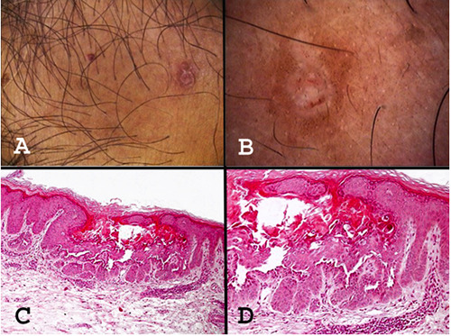
Warty Dyskeratoma (WD) is a rare condition consisting in single or multiple papular or nodular lesions of the skin or of the oral mucosamucosa. Histologically, a cupshaped epidermal invagination centred by a plug of epidermal hyperparakeratosis with suprabasal acantholysis and dyskeratosis is typically observed. A case of post-inflammatory WD, which was also observed by dermoscopy, is described. Dermoscopy showed an eight-shape whitish collarette surrounded by light brown pigmentation. A central white structureless area with an adjacent rosette were observed. Some small rust-coloured blood crusts were also observed in the centre of the lesion; no prominent vascular pattern was detected. The etiopathogenesis of this benign neoplasm could be multifactorial. Dermoscopy of WD is not specific but may help to ruling out other skin tumors.
Keywords: Warty dyskeratoma; dermatoscopy; dermoscopy; etiopathogenesis; histopathology.
©Copyright: the Author(s).
Conflict of interest statement
Conflict of interest: the authors declare no potential conflict of interests.
Figures
Similar articles
-
Warty dyskeratoma: The relationship between its dermoscopic and histopathological findings.Clin Case Rep. 2023 Jun 8;11(6):e7495. doi: 10.1002/ccr3.7495. eCollection 2023 Jun. Clin Case Rep. 2023. PMID: 37305894 Free PMC article.
-
Warty Dyskeratoma.2025 Apr 4. In: StatPearls [Internet]. Treasure Island (FL): StatPearls Publishing; 2025 Jan–. 2025 Apr 4. In: StatPearls [Internet]. Treasure Island (FL): StatPearls Publishing; 2025 Jan–. PMID: 40334037 Free Books & Documents.
-
Dermoscopy of Zosteriform and Swirling Pattern Type 1 Segmental Darier Disease.Acta Dermatovenerol Croat. 2022 Nov;30(3):191-193. Acta Dermatovenerol Croat. 2022. PMID: 36812281
-
Warty dyskeratoma/focal acantholytic dyskeratosis--an update on a rare oral lesion.J Oral Pathol Med. 2012 Mar;41(3):261-7. doi: 10.1111/j.1600-0714.2011.01082.x. Epub 2011 Sep 21. J Oral Pathol Med. 2012. PMID: 21936875 Review.
-
Oral Warty Dyskeratoma-A Systematic Review of the Literature.Diagnostics (Basel). 2022 May 20;12(5):1273. doi: 10.3390/diagnostics12051273. Diagnostics (Basel). 2022. PMID: 35626429 Free PMC article. Review.
Cited by
-
A Systematic Review of Diagnoses with Rosettes Under Dermoscopy.Dermatol Pract Concept. 2024 Apr 1;14(2):e2024125. doi: 10.5826/dpc.1402a125. Dermatol Pract Concept. 2024. PMID: 38810026 Free PMC article. Review.
-
Warty dyskeratoma: The relationship between its dermoscopic and histopathological findings.Clin Case Rep. 2023 Jun 8;11(6):e7495. doi: 10.1002/ccr3.7495. eCollection 2023 Jun. Clin Case Rep. 2023. PMID: 37305894 Free PMC article.
References
-
- Helwig EB. Proceedings of the 20th seminar on skin neoplasms and dermatoses, International Congress of Clinical Pathologists, 1954 Sept, Washington, DC, USA. Am Soc Clin Pathol 1955;53-6.
-
- Szymanski FJ. Warty dyskeratoma: a benign cutaneous tumor resembling Darier’s disease microscopically. Arch Dermatol 1957;75:567-72. - PubMed
-
- Graham JH, Helwig EB. Isolated dyskeratosis follicularis. AMA Arch Derm 1958;77:377-89 - PubMed
-
- Ackerman AB. Focal acantholytic dyskeratosis. Arch Dermatol 1972;106:702-6. - PubMed
-
- Diallo M, Cribier B, Scrivener Y. Warty dyskeratoma: infundibular histogenesis. Anatomoclinical study of 43 cases. Ann Dermatol Venereol 2007;134:633-6. - PubMed
LinkOut - more resources
Full Text Sources