. 2020 Dec;23(6):635-646.
doi: 10.4048/jbc.2020.23.e64.

In Vitro Analysis of Histology, Mechanics, and Safety of Radiation-free Pre-hydrated Human Acellular Dermal Matrix

Affiliations

In Vitro Analysis of Histology, Mechanics, and Safety of Radiation-free Pre-hydrated Human Acellular Dermal Matrix

Ji Young Kim et al. J Breast Cancer. 2020 Dec.

Abstract

Purpose: Acellular dermal matrix (ADM) supports tissue expanders or implants in implant-based breast reconstruction. The characteristics of ADM tissue are defined by the manufacturing procedure, such as decellularization, preservation, and sterilization, and are directly related to clinical outcomes. This study aimed to compare the properties of a new pre-hydrated-ADM (H-ADM-low) obtained using a decellularization reagent reduction process with a low concentration of detergent with those of radiation-sterilized H-ADM and freeze-dried ADM (FD-ADM).

Methods: ADMs were evaluated in terms of structure, mechanical quality, and cytotoxicity using histochemical staining, tensile strength testing, and in vitro cell viability analysis.

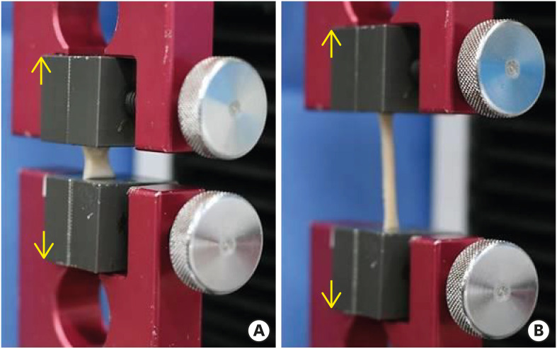
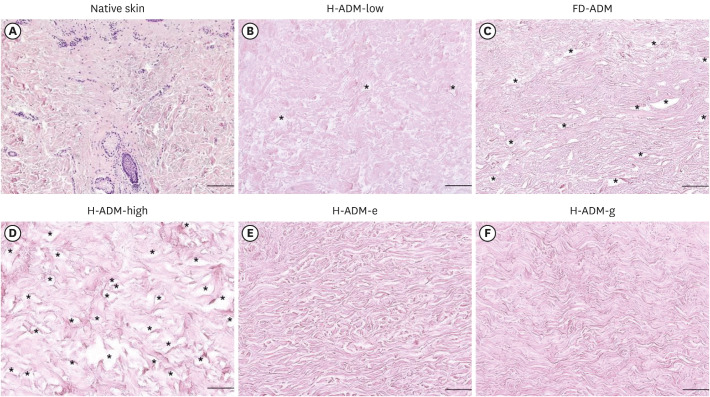
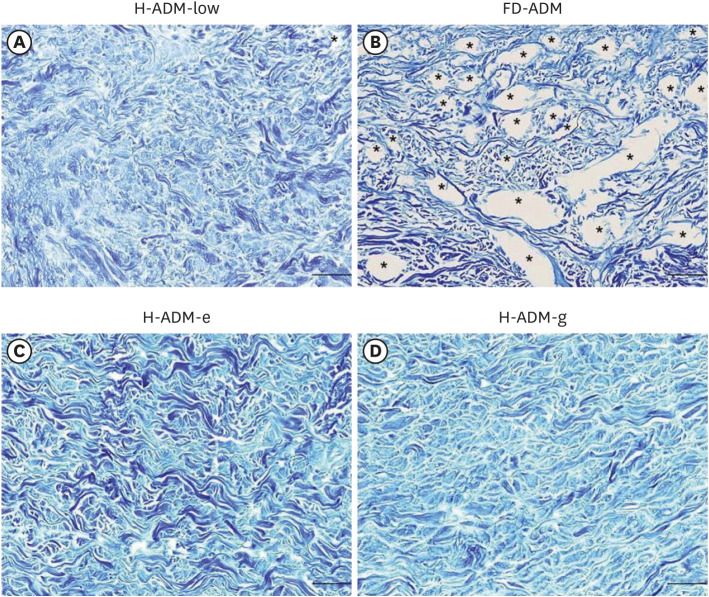
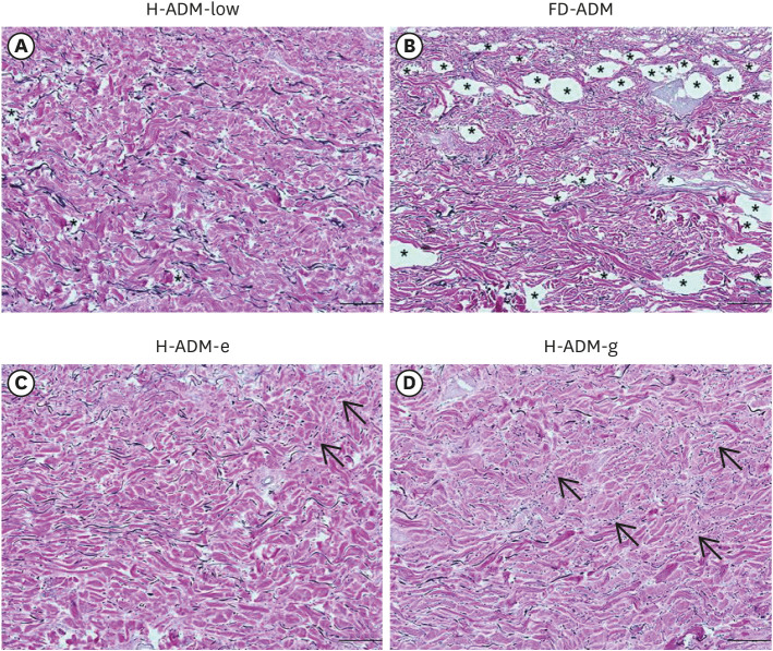
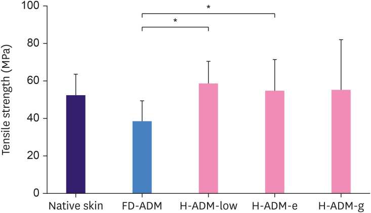
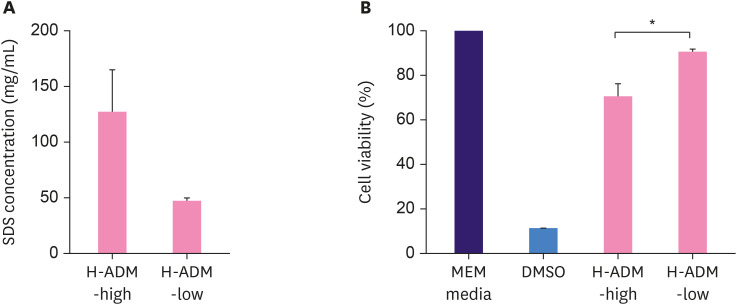
Results: The tissue structure of H-ADM-low (CGDERM ONE-STEP) was similar to that of native skin despite complete decellularization. By contrast, in FD-ADM, the tissue structure was damaged by the freeze-drying process, and radiation-sterilized H-ADM showed a compact fibrillar arrangement. Furthermore, matrix components such as collagen and elastin were preserved in H-ADM-low, whereas a loss of elastin fibers with fragmented distribution was observed in radiation-sterilized H-ADMs. H-ADM-low's tensile strength (58.84 MPa) was significantly greater than that of FD-ADM (38.60 MPa) and comparable with that of radiation-sterilized H-ADMs. The residual detergent content in H-ADM-low (47.45 mg/L) was 2.67-fold lower than that of H-ADM decellularized with a conventional detergent concentration (126.99 mg/mL), and this finding was consistent with the cell viability results (90.7% and 70.7%, respectively), indicating that H-ADM-low has very low cytotoxicity.

Conclusions: H-ADM-low produced through aseptic processes retains the original tissue structure, demonstrates excellent mechanical properties, and does not affect cell viability. Therefore, this newer H-ADM is suitable for use in implant-based breast reconstruction.

Keywords: Acellular dermis; Biological preservation; Breast implantation; Detergents; In vitro techniques.


Conflict of interest statement

Conflict of Interest: The authors declare that they have no competing interests.

Figures

Figure 1
Figure 1. Tensile strength testing. (A) The tissue was fixed in the universal testing machine and (B) stretched in the direction of the yellow arrows.
Figure 2
Figure 2. Evaluation of the structure and decellularization status in ADM samples. Samples were fixed and stained with HE; representative images are shown (magnification × 100; scale bars, 200 μm). (A) Native skin, (B) H-ADM-low, (C) FD-ADM, (D) H-ADM-high, (E) H-ADM-e, and (F) H-ADM-g. The cell nuclei are stained dark-blue or purple and the extracellular material, such as collagen, is stained pink. The stars indicate the location of the void.
ADM = acellular dermal matrix; HE = hematoxylin and eosin; Native skin = unprocessed human cellular dermal matrix; H-ADM-low = hydrated-acellular dermal matrix prepared using low sodium dodecyl sulfate concentration; FD-ADM = freeze-dried acellular dermal matrix; H-ADM-high = hydrated-acellular dermal matrix prepared using conventional high SDS concentration; H-ADM-e = E-beam radiation-sterilized pre-hydrated acellular dermal matrix; H-ADM-g = gamma radiation-sterilized pre-hydrated acellular dermal matrix.
Figure 3
Figure 3. Collagen structure in ADM samples. Representative images of tissues stained with MT are shown (magnification × 100; scale bars, 200 μm) (A) H-ADM-low, (B) FD-ADM, (C) H-ADM-e, and (D) H-ADM-g. The collagen fibers are stained blue. The stars indicate the location of the void.
ADM = acellular dermal matrix; MT = Masson's trichrome; H-ADM-low = hydrated-acellular dermal matrix prepared using low sodium dodecyl sulfate concentration; FD-ADM = freeze-dried acellular dermal matrix; H-ADM-e = E-beam radiation-sterilized pre-hydrated acellular dermal matrix; H-ADM-g = gamma radiation-sterilized pre-hydrated acellular dermal matrix.
Figure 4
Figure 4. Elastin structure in ADM samples. Representative images of tissues stained with VVG staining method are shown (magnification × 100; scale bars, 200 μm). (A) H-ADM-low, (B) FD-ADM, (C) H-ADM-e, (D) H-ADM-g. The elastic fibers and collagen fibers are stained black and red, respectively. The arrows indicate the fragmented elastic fibers and the stars indicate the location of the void.
ADM = acellular dermal matrix; VVG = Verhoeff-van Gieson; H-ADM = pre-hydrated acellular dermal matrix; H-ADM-low = hydrated-acellular dermal matrix prepared using low sodium dodecyl sulfate concentration; FD-ADM = freeze-dried acellular dermal matrix; H-ADM-e = E-beam radiation-sterilized pre-hydrated acellular dermal matrix; H-ADM-g = gamma radiation-sterilized pre-hydrated acellular dermal matrix.
Figure 5
Figure 5. Evaluation of tissue mechanical properties by the tensile strength test. The results are presented as the mean ± standard deviation (n = 5).
Native skin = unprocessed human cellular dermal matrix; FD-ADM = freeze-dried acellular dermal matrix; H-ADM-low = hydrated-ADM prepared using sodium dodecyl sulfate concentration, H-ADM-e = E-beam radiation-sterilized pre-hydrated acellular dermal matrix; H-ADM-g = gamma radiation-sterilized pre-hydrated acellular dermal matrix. *p < 0.05.
Figure 6
Figure 6. Residual detergent concentration and cytotoxicity of H-ADM prepared using different concentrations of detergent. (A) H-ADM-high and H-ADM-low samples were decellularized using high and low SDS concentrations, respectively. The residual detergent concentrations (mg/mL) are presented as the mean ± SD (n = 3). (B) L929 cells were treated with MEM media (control), DMSO (which is toxic to cells), and eluates of H-ADM-high and H-ADM-low. The cytotoxicity results (%) are presented as the mean ± SD (n = 3).
H-ADM = pre-hydrated acellular dermal matrix; H-ADM-high = hydrated-acellular dermal matrix prepared using high sodium dodecyl sulfate concentration; H-ADM-low = hydrated-acellular dermal matrix prepared using low sodium dodecyl sulfate concentration; SDS = sodium dodecyl sulfate; MEM = minimum essential medium; DMSO = dimethyl sulfoxide; FD-ADM = freeze-dried acellular dermal matrix; H-ADM-e = E-beam radiation-sterilized pre-hydrated acellular dermal matrix; H-ADM-g = gamma radiation-sterilized pre-hydrated acellular dermal matrix; SD = standard deviation. *p < 0.05.

Similar articles

Cited by

References

    1. Capito AE, Tholpady SS, Agrawal H, Drake DB, Katz AJ. Evaluation of host tissue integration, revascularization, and cellular infiltration within various dermal substrates. Ann Plast Surg. 2012;68:495–500. - PubMed
    1. Garcia O, Jr, Scott JR. Analysis of acellular dermal matrix integration and revascularization following tissue expander breast reconstruction in a clinically relevant large-animal model. Plast Reconstr Surg. 2013;131:741e–751e. - PubMed
    1. Bellows CF, Alder A, Helton WS. Abdominal wall reconstruction using biological tissue grafts: present status and future opportunities. Expert Rev Med Devices. 2006;3:657–675. - PubMed
    1. Galili U. The α-Gal epitope (Galα1-3Galβ1-4GlcNAc-R) in xenotransplantation. Biochimie. 2001;83:557–563. - PubMed
    1. Armour AD, Fish JS, Woodhouse KA, Semple JL. A comparison of human and porcine acellularized dermis: interactions with human fibroblasts in vitro . Plast Reconstr Surg. 2006;117:845–856. - PubMed