

Intramedullary spinal schistosomiasis in a child with acute myelopathy: A case report
- PMID: 33408905
- PMCID: PMC7771502
- DOI: 10.25259/SNI_484_2020
Intramedullary spinal schistosomiasis in a child with acute myelopathy: A case report
Abstract
Background: Neuroschistosomiasis is defined as an infection of the nervous system caused by Schistosoma mansoni. Neuroschistosomiasis is an important differential diagnostic consideration in pediatric patients presenting with myelopathy. Surgical excision combined with antiparasitic drugs typically provides a satisfactory outcome and often results in neurological recovery.
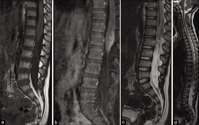
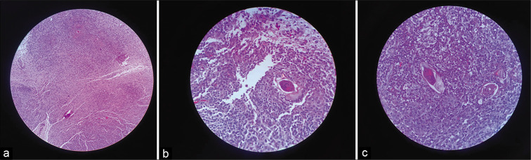
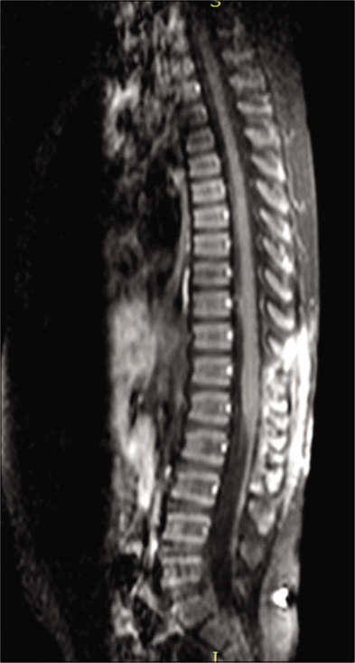
Case description: A 4-year-old child presented with acute and progressive myelopathy. A thoracolumbar magnetic resonance image revealed a T12-L2 conus medullaris mass that was isointense on T1 and hyperintense on T2 (with an extensive syringomyelia at the thoracic spinal cord) and showed enhanced heterogeneity with gadolinium. The lesion was excised through T12-L2 laminotomy. Intraoperatively, the tumor appeared reddish and infiltrative. The frozen section suggested a granulomatous process, while the final pathology confirmed conus medullaris schistosomiasis.
Conclusion: Schistosomal myeloradiculopathy should be considered among the different diagnosis in children presenting with lower thoracic region, conus medullaris, and/or cauda equina infiltrative spinal masses.
Keywords: Conus medullaris; Myelopathy; Neuroschistosomiasis; Schistosomiasis; Spinal.
Copyright: © 2020 Surgical Neurology International.
Conflict of interest statement
There are no conflicts of interest.
Figures
References
-
- Camargo ST, Dantas FR, Teixeira AL. Schistosomal myelopathy mimicking spinal cord neoplasm. Scand J Infect Dis. 2005;37:365–98. - PubMed
-
- Kim AH, Maher CO, Smith ER. Lumbar intramedullary spinal schistosomiasis presenting as progressive paraparesis: Case report. Neurosurgery. 2006;58:E996. - PubMed
-
- Labeodan AO, Sur M. Intramedullary schistosomiasis. Pediatr Neurosurg. 2003;39:14–6. - PubMed
-
- Lighter J, Kim M, Krasinski K. Intramedullary schistosomiasis presenting in an adolescent with prolonged intermittent back pain. Pediatr Neurol. 2008;39:44–7. - PubMed
Publication types
LinkOut - more resources
Full Text Sources