

Sphenoid wing dural arteriovenous fistula: A case report and literature review
- PMID: 33408924
- PMCID: PMC7771485
- DOI: 10.25259/SNI_571_2020
Sphenoid wing dural arteriovenous fistula: A case report and literature review
Abstract
Background: Sphenoid wing dural arteriovenous fistula (SWDAVF) is rare that is typically fed by middle meningeal artery feeders and that drain through the sphenoparietal sinus or middle cerebral vein. Here, we report a case of SWDAVF treated by coils placed in the venous aneurysm through the contralateral cavernous sinus (CS).
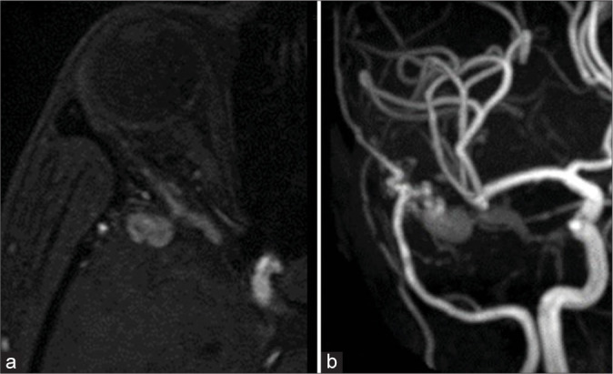
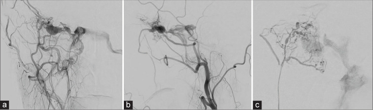
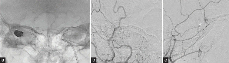
Case description: A 37-year-old woman was admitted to our hospital with headache and bilateral oculomotor nerve palsy. Magnetic resonance images and an angiogram showed a venous aneurysm in the right middle cranial fossa. A DAVF, consisting of two main feeders, was diagnosed based on the angiogram findings. The fistula drained into the left inferior petrosal sinus (IPS) through the left CS and right IPS. Given the remarkable extent of venous ectasia together with the headache and right abducens nerve paralysis, endovascular treatment was initiated. A transvenous approach through the right IPS was not feasible, as it is strenuous to insert the microcatheter into the right IPS. Thus, we tried an approach through the left IPS. The venous aneurysm was embolized with coils. The postoperative course was uneventful, and postoperative cerebral angiography confirmed disappearance of the fistula.
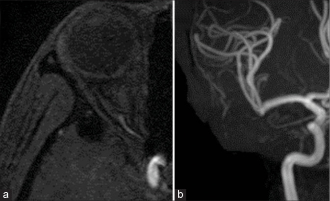
Conclusion: A SWDAVF is extremely rare. In our case, since the AVF drained into the contralateral CS, contralateral ocular symptoms occurred. Endovascular occlusion of the venous aneurysm and fistula was achieved through a transvenous approach.
Keywords: Cavernous sinus; Dural arteriovenous fistula; Endovascular; Venous aneurysm.
Copyright: © 2020 Surgical Neurology International.
Conflict of interest statement
There are no conflicts of interest.
Figures
Similar articles
-
Transarterial Sinus Embolization for a Dural Arteriovenous Fistula in a Sinus of the Lesser Sphenoid Wing: A Case Report.NMC Case Rep J. 2017 Mar 7;4(2):47-50. doi: 10.2176/nmccrj.cr.2016-0076. eCollection 2017 Apr. NMC Case Rep J. 2017. PMID: 28664026 Free PMC article.
-
Transvenous embolization with a combination of detachable coils and Onyx for a complicated cavernous dural arteriovenous fistula.Chin Med J (Engl). 2008 Sep 5;121(17):1651-5. Chin Med J (Engl). 2008. PMID: 19024093
-
Cortical Venous Approach for Transvenous Embolization of a Greater Sphenoid Wing Dural Arteriovenous Fistula: A Case Report.J Neuroendovasc Ther. 2025;19(1):2024-0071. doi: 10.5797/jnet.cr.2024-0071. Epub 2024 Oct 31. J Neuroendovasc Ther. 2025. PMID: 40018287 Free PMC article.
-
Transvenous occlusion of dural cavernous sinus fistulas through the thrombosed inferior petrosal sinus: report of four cases and review of the literature.Surg Neurol. 2000 Jul;54(1):42-54. doi: 10.1016/s0090-3019(00)00260-3. Surg Neurol. 2000. PMID: 11024506 Review.
-
Intra-arterial onyx embolisation of sphenobasilar sinus fistula using pressure cooker technique: case report and review of the literature.Neuroradiol J. 2021 Apr;34(2):131-134. doi: 10.1177/1971400920972512. Epub 2020 Nov 11. Neuroradiol J. 2021. PMID: 33176554 Free PMC article. Review.
Cited by
-
Sphenoid Wing Dural Arteriovenous Fistulas.J Neuroendovasc Ther. 2025;19(1):2023-0034. doi: 10.5797/jnet.ra.2023-0034. Epub 2023 Dec 23. J Neuroendovasc Ther. 2025. PMID: 40018285 Free PMC article. Review.
-
Trans-Galen Approach for Embolization of Sphenoparietal Sinus Dural Arteriovenous Fistulas.Neurointervention. 2025 Jul;20(2):114-118. doi: 10.5469/neuroint.2025.00094. Epub 2025 Mar 12. Neurointervention. 2025. PMID: 40068630 Free PMC article.
-
Spontaneous obliteration of a greater sphenoid wing dural arteriovenous fistula involving the diploic venous system.Surg Neurol Int. 2025 Mar 21;16:99. doi: 10.25259/SNI_1113_2024. eCollection 2025. Surg Neurol Int. 2025. PMID: 40206771 Free PMC article.
-
Iatrogenic mixed pial and dural arteriovenous fistula after pterional approach for surgical clipping of aneurysm: A case report.J Cerebrovasc Endovasc Neurosurg. 2023 Dec;25(4):440-446. doi: 10.7461/jcen.2023.E2022.12.004. Epub 2023 May 16. J Cerebrovasc Endovasc Neurosurg. 2023. PMID: 37189252 Free PMC article.
-
Non-aggressive, sinus-type greater sphenoid wing dural arteriovenous fistula with shunt point in the laterocavernous sinus mimicking a cavernous sinus dural arteriovenous fistula converted to aggressive, non-sinus-type.BMJ Case Rep. 2025 Apr 24;18(4):e265715. doi: 10.1136/bcr-2025-265715. BMJ Case Rep. 2025. PMID: 40280581 Free PMC article.
References
-
- Awad IA, Little JR, Akarawi WP, Ahl J. Intracranial dural arteriovenous malformations: Factors predisposing to an aggressive neurological course. J Neurosurg. 1990;72:839–50. - PubMed
-
- Ezura M, Takahashi A, Mizoi K. Dural arteriovenous shunts involving the sphenoparietal sinus: A case report. Interv Neuroradiol. 1996;2:223–8. - PubMed
-
- Fukuda H, Miyake K, Kunieda T, Murao K. Endovascular treatment of sphenoid wing dural arteriovenous fistula with pure cortical venous drainage. J Stroke Cerebrovasc Dis. 2014;23:1730–5. - PubMed
-
- Khadavi NM, Mancini R, Nakra T. Rare dural arteriovenous fistula of the lesser sphenoid wing sinus. Ophthalmic Plast Reconstr Surg. 2009;25:404–6. - PubMed
Publication types
LinkOut - more resources
Full Text Sources