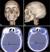
99mTc-UBI 29-41 bone SPECT/CT scan in craniofacial Actinomyces israelii: Misdiagnosis of cranial bone tumor - A case report
- PMID: 33408927
- PMCID: PMC7771506
- DOI: 10.25259/SNI_684_2020
99mTc-UBI 29-41 bone SPECT/CT scan in craniofacial Actinomyces israelii: Misdiagnosis of cranial bone tumor - A case report
Abstract
Background: Actinomycosis is a rare infection, frequently misdiagnosed as a neoplasia. This chronic and granulomatous disease is caused by Actinomyces israelii species. Cervicofacial actinomycosis occurs in 60% of cases and the diagnosis is commonly made by histopathology study.
Case description: We report a case of fronto-orbital osteomyelitis initially misdiagnosed as a cranial bone meningioma, but later proved to be a case of actinomycosis. 99mTechnetium (99mTc) three-phase bone single-photon emission computed tomography/computed tomography (SPECT/CT) and 99mTc-ubiquicidin (UBI) 29-41 bone SPECT/CT scans were performed to corroborate the control of the infection.
Conclusion: Craniofacial actinomycosis is the most common presentation of actinomycosis. However, it continues to be a rare and difficult disease to diagnose and is often confused with a neoplastic process. The 99mTc-UBI 29-41 bone SPECT/CT scan could be an auxiliary noninvasive diagnostic alternative and a follow-up method for these patients.
Keywords: Actinomyces; Craniofacial; Osteomyelitis.
Copyright: © 2020 Surgical Neurology International.
Conflict of interest statement
There are no conflicts of interest.
Figures


Similar articles
-
Utility of ⁹⁹mTc-labelled antimicrobial peptide ubiquicidin (29-41) in the diagnosis of diabetic foot infection.Eur J Nucl Med Mol Imaging. 2013 May;40(5):737-43. doi: 10.1007/s00259-012-2327-1. Epub 2013 Jan 30. Eur J Nucl Med Mol Imaging. 2013. PMID: 23361858 Clinical Trial.
-
99mTechnetium-Ubiquicidin Scan with Single-Photon Emission Computed Tomography/Computed Tomography in Skull Base Osteomyelitis.Indian J Nucl Med. 2023 Jul-Sep;38(3):297-300. doi: 10.4103/ijnm.ijnm_192_22. Epub 2023 Oct 10. Indian J Nucl Med. 2023. PMID: 38046968 Free PMC article.
-
Clinical utility of 99mTc-ubiquicidin (29-41) as an adjunct to bone scan in differentiating infected versus noninfected loosening of prosthesis before revision surgery.Nucl Med Commun. 2017 Apr;38(4):285-290. doi: 10.1097/MNM.0000000000000648. Nucl Med Commun. 2017. PMID: 28244975
-
Clinical Applications of Technetium-99m Quantitative Single-Photon Emission Computed Tomography/Computed Tomography.Nucl Med Mol Imaging. 2019 Jun;53(3):172-181. doi: 10.1007/s13139-019-00588-9. Epub 2019 Mar 15. Nucl Med Mol Imaging. 2019. PMID: 31231437 Free PMC article. Review.
-
Atypical Form of Cervicofacial Actinomycosis Involving the Skull Base and Temporal Bone.Ann Otol Rhinol Laryngol. 2019 Feb;128(2):152-156. doi: 10.1177/0003489418808541. Epub 2018 Oct 29. Ann Otol Rhinol Laryngol. 2019. PMID: 30371104 Review.
References
Publication types
LinkOut - more resources
Full Text Sources
Molecular Biology Databases
