. 2020 Nov 10;11(12):7062-7078.
doi: 10.1364/BOE.408649. eCollection 2020 Dec 1.

Highly sensitive detection of cytochrome c in the NSCLC serum using a hydrophobic paper based-gold nanourchin substrate

Affiliations

Highly sensitive detection of cytochrome c in the NSCLC serum using a hydrophobic paper based-gold nanourchin substrate

Yue Sun et al. Biomed Opt Express. .

Abstract

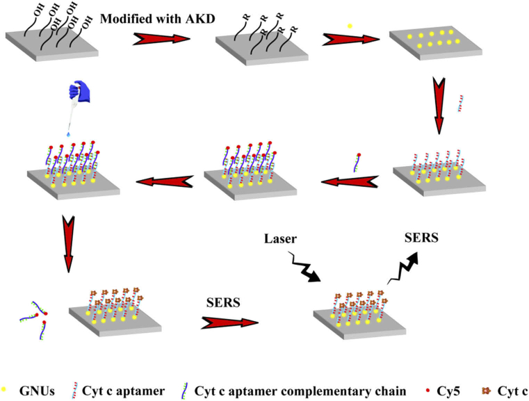
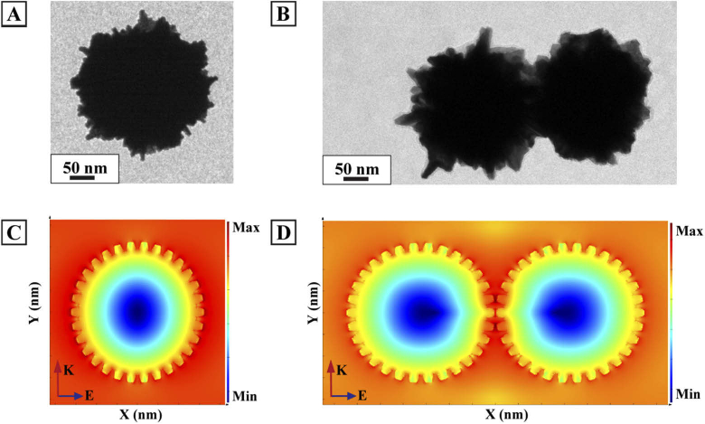
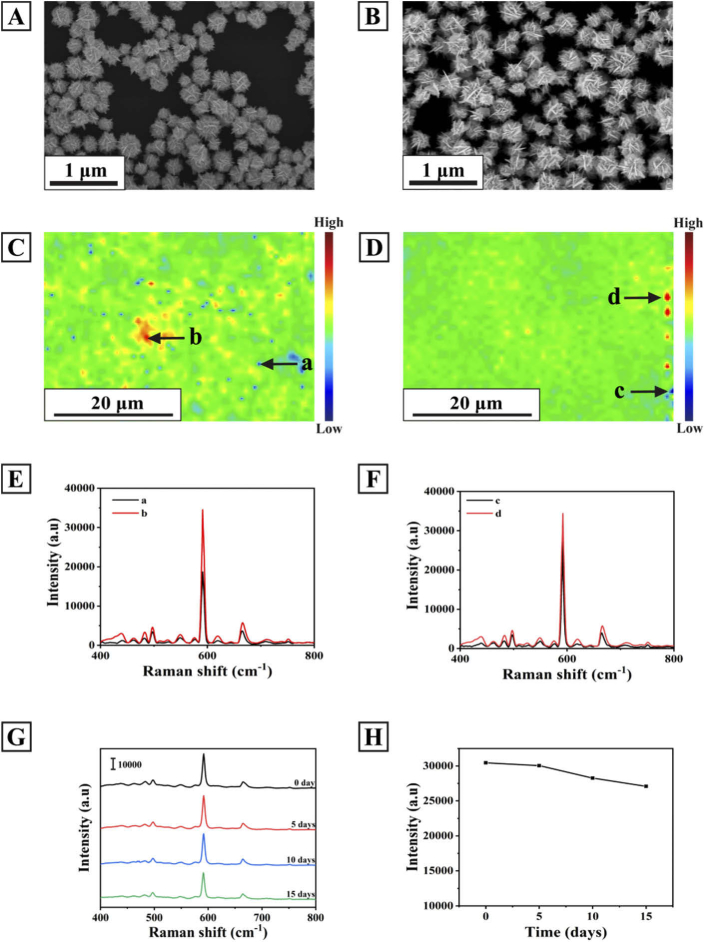
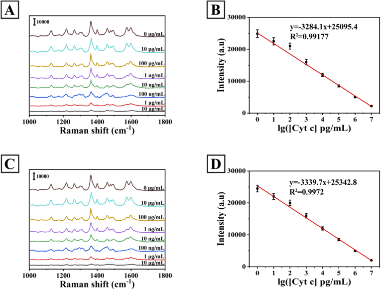
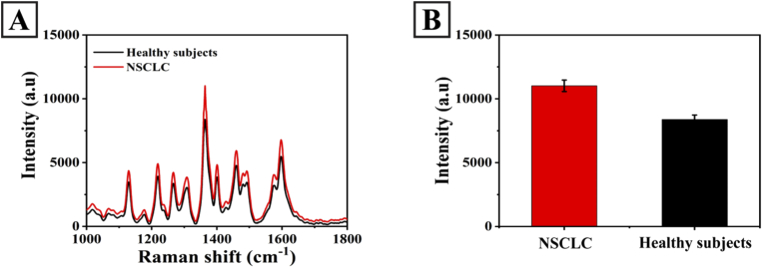
Cytochrome c (Cyt c) is a biomarker of early apoptosis that plays a critical role in the diagnosis and therapy of non-small cell lung cancer (NSCLC). In this work, we proposed a novel surface-enhanced Raman scattering (SERS)-based biosensor to implement the ultrasensitive detection of Cyt c in the serum of NSCLC patients. The SERS-supporting substrates based on hydrophobic filter paper were composed of gold nanourchins (GNUs) surface-functionalized with the Cyt c aptamer and the cyanine 5-labeled complementary DNA. In the existence of Cyt c, it could specifically bind to its aptamer, which leads to the detachment of complementary strands modified with Cy5 and the great weakness of SERS signal. The finite-difference time domain (FDTD) simulation showed that the excellent SERS performance of GNUs aggregation was strongly dependent on a large number of "hot spots" at the tips and between the nanogaps of aggregated GNUs. Alkyl ketene dimer (AKD) was used to make the filter paper modify its property from hydrophilic to hydrophobic, which consequently increased the density of GNUs and extended the retention time of the analyte. SERS biosensors based on hydrophobic paper exhibited prominent reproducibility and selectivity. The detection limit of Cyt c in PBS was 1.148 pg/mL, while the detection limit in human serum was 1.79 pg/mL. Moreover, the analysis of the serum samples of healthy subjects and NSCLC patients confirmed the feasibility of its clinical application. The results were consistent with enzyme-linked immunosorbent assay results. This method can be a powerful strategy for quantitative detection of extracellular Cyt c, and it is expected that the SERS-based biosensors could be applied in the practical clinical diagnoses of NSCLC.


Conflict of interest statement

The authors declare that there are no conflicts of interest related to this article.

Figures

Fig. 1.
Fig. 1.
Schematic illustration of Cyt c detection mechanism with GNUs biosensors based on SERS.
Fig. 2.
Fig. 2.
Typical SEM (A, B) and TEM (C) images of the GNUs. (D) The HRTEM image of the GNUs. (E) The SAED pattern of the GNUs. (F) The representative UV-visible absorption spectrum of the GNUs. The insert shows the solution of the GNUs.
Fig. 3.
Fig. 3.
TEM images of (A) a single GNU, (B) GNUs dimer. The corresponding simulated electric field distribution images of (C) a single GNU, (D) GNUs dimer.
Fig. 4.
Fig. 4.
SEM images of (A) the GNUs-modified paper substrate and (B) the GNUs-modified h-paper substrate. SERS mapping of NBA (5×10⁻⁶ M) measured at 592 cm-1 using (C) the GNUs-modified paper substrate and (D) the GNUs-modified h-paper substrate. (E) The SERS spectra at the strongest point (Fig. 4(C)-(b)) and the weakest point (Fig. 4(C)-(a)) on the GNUs-modified paper substrate. (F) The SERS spectra at the strongest point (Fig. 4(D)-(d)) and the weakest point (Fig. 4(D)-(c)) on the GNUs-modified h-paper substrate. (G) The average SERS spectra of the NBA-labeled GNUs-modified h-paper substrate after the storage of 0 day, 5 days, 10 days, 15 days at room temperature. (H) SERS intensities of the bands at 592 cm-1 corresponding to the SERS spectra (G).
Fig. 5.
Fig. 5.
(A) SERS spectra of the GNUs substrate (curve a), the GNUs substrate with Cyt c aptamer modification (curve b), the GNUs substrate with aptamer and Cy5-labeled complementary DNA (curve c), the SERS biosensor in the presence of Cyt c (curve d). (B) The corresponding intensities bar graphs of characteristic peaks of Cy5 at 1360 cm-1.
Fig. 6.
Fig. 6.
(A) SERS spectra measured for the detection of Cyt c, glucose (Glu), immunization globulin G (lgG), human serum albumin (HSA), bovine serum albumin (BSA) and ascorbic acid (AA) at the same concentration of 10 μg/mL in PBS. (B) SERS intensities of Cy5 characteristic peak at 1360 cm-1 corresponding to A. (C) The reproducibility of SERS biosensor platform. (D) SERS intensities of Cy5 characteristic peak at 1360 cm-1 corresponding to C.
Fig. 7.
Fig. 7.
(A) SERS spectra of Cy5 for quantitative evaluation of the Cyt c with different concentrations from 0 to 10 μg/mL in PBS buffer. (B) Calibration plot of the changed SERS signal intensities (1360 cm-1) versus the logarithm of Cyt c concentrations corresponding to A. (C) SERS spectra of Cy5 for quantitative evaluation of the Cyt c with different concentrations from 0 to 10 μg/mL in the serum. (D) Calibration plot of the changed SERS signal intensities (1360 cm-1) versus the logarithm of Cyt c concentrations corresponding to C.
Fig. 8.
Fig. 8.
(A) The mean SERS spectra of different clinical serum samples. (B) The bar charts of the intensities of the peaks at 1360 cm-1 corresponding to A.

Similar articles

Cited by

References

    1. Parkin D. M., Bray F., Ferlay J., Pisani P., “Global cancer statistics, 2002,” Ca-Cancer J. Clin. 55(2), 74–108 (2005).10.3322/canjclin.55.2.74 - DOI - PubMed
    1. Asamura H., Goya T., Koshiishi Y., Sohara Y., Eguchi K., Mori K., Nakanishi Y., Tsuchiya R., Shimokata K., Inoue H., Nitkiwa T., Miyaoka E., “A Japanese lung cancer registry study-Prognosis of 13,010 resected lung cancers,” J. Thorac. Oncol. 3(1), 46–52 (2008).10.1097/JTO.0b013e31815e8577 - DOI - PubMed
    1. Haaland B., Tan P. S., de Castro G. J., Lopes G., “Metaanalysis of first-line therapies in advanced non-small-cell lung cancer harboring EGFR-activating mutations,” J. Thorac. Oncol. 9(6), 805–811 (2014).10.1097/JTO.0000000000000156 - DOI - PMC - PubMed
    1. Bray F., Ferlay J., Soerjomataram I., Siegel R. L., Torre L. A., Jemal A., “Global cancer statistics 2018: GLOBOCAN estimates of incidence and mortality worldwide for 36 cancers in 185 countries,” Ca-Cancer J. Clin. 68(6), 394–424 (2018).10.3322/caac.21492 - DOI - PubMed
    1. Amouzadeh Tabrizi M., Ferre-Borrull J., Marsal L. F., “Highly sensitive IRS based biosensor for the determination of cytochrome c as a cancer marker by using nanoporous anodic alumina modified with trypsin,” Biosens. Bioelectron. 149, 111828 (2020).10.1016/j.bios.2019.111828 - DOI - PubMed

LinkOut - more resources