

Multiple-pulse damage thresholds of retinal explants in the ns-time regime
- PMID: 33408997
- PMCID: PMC7747910
- DOI: 10.1364/BOE.412012
Multiple-pulse damage thresholds of retinal explants in the ns-time regime
Abstract
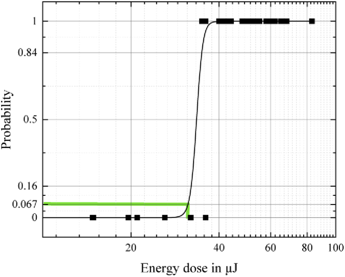
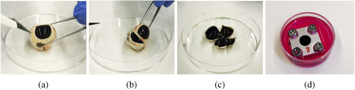
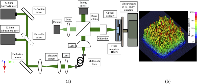
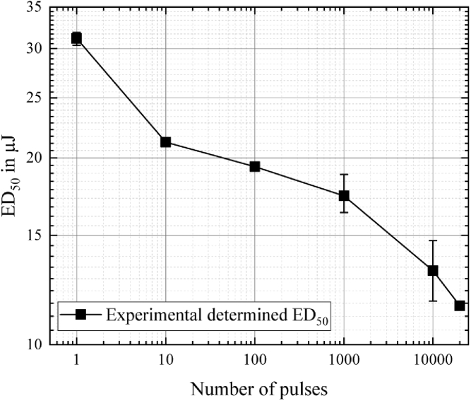
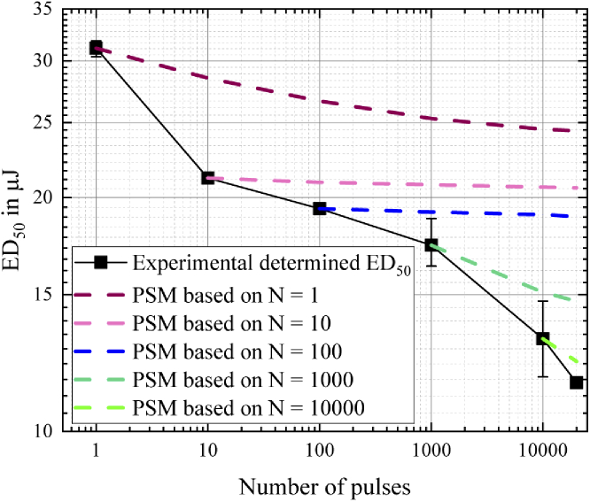
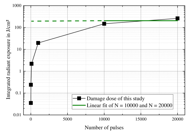
The data situation of laser-induced damage measurements after multiple-pulse irradiation in the ns-time regime is limited. Since the laser safety standard is based on damage experiments, it is crucial to determine damage thresholds. For a better understanding of the underlying damage mechanism after repetitive irradiation, we generate damage thresholds for pulse sequences up to N = 20 000 with 1.8 ns-pulses using a square-core fiber and a pulsed Nd:YAG laser. Porcine retinal pigment epithelial layers were used as tissue samples, irradiated with six pulse sequences and evaluated for damage by fluorescence microscopy. The damage thresholds decreased from 31.16 µJ for N = 1 to 11.56 µJ for N = 20 000. The reduction indicates photo-chemical damage mechanisms after reaching a critical energy dose.
© 2020 Optical Society of America under the terms of the OSA Open Access Publishing Agreement.
Conflict of interest statement
The authors declare that there are no conflicts of interest related to this article.
Figures
Similar articles
-
Threshold determinations for selective retinal pigment epithelium damage with repetitive pulsed microsecond laser systems in rabbits.Ophthalmic Surg Lasers. 2002 Sep-Oct;33(5):400-9. Ophthalmic Surg Lasers. 2002. PMID: 12358294
-
Ablation of porcine ligamentum flavum with Ho:YAG, q-switched Ho:YAG, and quadrupled Nd:YAG lasers.Lasers Surg Med. 2015 Dec;47(10):839-51. doi: 10.1002/lsm.22424. Epub 2015 Sep 28. Lasers Surg Med. 2015. PMID: 26415136 Free PMC article.
-
Bubble formation as primary interaction mechanism in retinal laser exposure with 200-ns laser pulses.Lasers Surg Med. 1998;22(4):240-8. doi: 10.1002/(sici)1096-9101(1998)22:4<240::aid-lsm9>3.0.co;2-p. Lasers Surg Med. 1998. PMID: 9603286
-
Review of thresholds and recommendations for revised exposure limits for laser and optical radiation for thermally induced retinal injury.Health Phys. 2011 Feb;100(2):210-20. doi: 10.1097/HP.0b013e3181ea51e3. Health Phys. 2011. PMID: 21399437 Review.
-
Capabilities and limitations of a new thermal finite volume model for the evaluation of laser-induced thermo-mechanical retinal damage.Comput Biol Med. 2020 Jul;122:103835. doi: 10.1016/j.compbiomed.2020.103835. Epub 2020 May 25. Comput Biol Med. 2020. PMID: 32479348 Review.
Cited by
-
Nanosecond multipulse retinal damage thresholds of elongated irradiance profiles in explant measurements and simulations.J Biomed Opt. 2023 Dec;28(12):125001. doi: 10.1117/1.JBO.28.12.125001. Epub 2023 Dec 2. J Biomed Opt. 2023. PMID: 38074214 Free PMC article.
References
-
- IEC 60825-1 , Safety of Laser Products - Part 1: Equipment Classification and Requirements (International Electrotechnical Commission, Geneva, 2014), 3rd ed.
-
- Schulmeister K., Jean M., “Manifestation of the strong non-linearity of thermal injury,” in International Laser Safety Conference, vol. 2011 (LIA, 2011), pp. 201–204.
-
- Birngruber R., Hillenkamp F., Gabel V., “Theoretical investigations of laser thermal retinal injury,” Health Phys. 48(6), 781–796 (1985). - PubMed
LinkOut - more resources
Full Text Sources
Miscellaneous