. 2020 Dec 21:8:597423.
doi: 10.3389/fcell.2020.597423. eCollection 2020.

Reversal of Endothelial Extracellular Vesicle-Induced Smooth Muscle Phenotype Transition by Hypercholesterolemia Stimulation: Role of NLRP3 Inflammasome Activation

Affiliations

Reversal of Endothelial Extracellular Vesicle-Induced Smooth Muscle Phenotype Transition by Hypercholesterolemia Stimulation: Role of NLRP3 Inflammasome Activation

Xinxu Yuan et al. Front Cell Dev Biol. .

Abstract

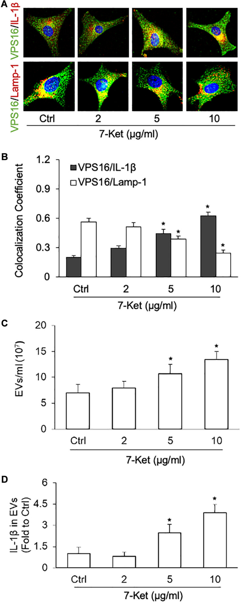
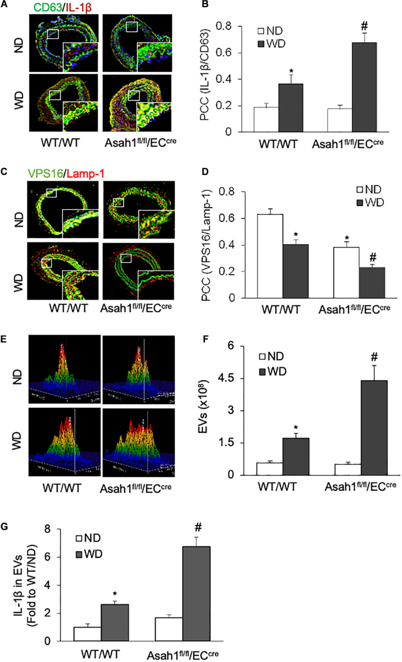
Recent studies reported that vascular endothelial cells (ECs) secrete NLR family pyrin domain-containing 3 (NLRP3) inflammasome products such as interleukin-1β (IL-1β) via extracellular vesicles (EVs) under various pathological conditions. EVs represent one of the critical mechanisms mediating the cell-to-cell communication between ECs and vascular smooth muscle cells (VSMCs). However, whether or not the inflammasome-dependent EVs directly participate in the regulation of VSMC function remains unknown. In the present study, we found that in cultured carotid ECs, atherogenic stimulation by oxysterol 7-ketocholesterol (7-Ket) induced NLRP3 inflammasome formation and activation, reduced lysosome-multivesicular bodies (MVBs) fusion, and increased secretion of EVs that contain inflammasome product IL-1β. These EC-derived IL-1β-containing EVs promoted synthetic phenotype transition of co-cultured VSMCs, whereas EVs from unstimulated ECs have the opposite effects. Moreover, acid ceramidase (AC) deficiency or lysosome inhibition further exaggerated the 7-Ket-induced release of IL-1β-containing EVs in ECs. Using a Western diet (WD)-induced hypercholesterolemia mouse model, we found that endothelial-specific AC gene knockout mice (Asah1fl/fl/ECCre) exhibited augmented WD-induced EV secretion with IL-1β and more significantly decreased the interaction of MVBs with lysosomes in the carotid arterial wall compared to their wild-type littermates (WT/WT). The endothelial AC deficiency in Asah1fl/fl/ECCre mice also resulted in enhanced VSMC phenotype transition and accelerated neointima formation. Together, these results suggest that NLRP3 inflammasome-dependent IL-1β production during hypercholesterolemia promotes VSMC phenotype transition to synthetic status via EV machinery, which is controlled by lysosomal AC activity. Our findings provide novel mechanistic insights into understanding the pathogenic role of endothelial NLRP3 inflammasome in vascular injury through EV-mediated EC-to-VSMC regulation.

Keywords: acid ceramidase; ceramide; endothelial cells; extracellular vesicles; lysosome.


Conflict of interest statement

The authors declare that the research was conducted in the absence of any commercial or financial relationships that could be construed as a potential conflict of interest.

Figures

FIGURE 1
FIGURE 1
Phenotype transition of vascular smooth muscle cells (VSMCs) cocultured with extracellular vesicles (EVs) isolated from the primary cultured carotid arterial endothelial cells (ECs). Primary cultured carotid arterial ECs were treated with 7-Ketocholesterol (7-Ket) (0-10 μg/ml) for 24 h. (A,C) Representative Western blot gel documents showing the expression of vimentin and SM22α induced by EVs collected from the carotid arterial ECs with (EVs-7-Ket) or without 7-Ket treatment (EVs-Ctrl). (B,D) The summarized data showing the ratio of vimentin with SM22α protein. (E) Representative wound healing assay images presenting the (1,3,5 × 109 EVs) effects of EVs on CAMs migration. (F) Summarized data showing the dose effects of EVs on CAMs migration. (G) Summarized data showing the dose effects of EVs on CAMs proliferation. Data are expressed as means ± SEM, n = 5. *p < 0.05 vs. Ctrl group.
FIGURE 2
FIGURE 2
NLRP3 inflammasome formation and activation dose-dependently stimulated by 7-Ket in the primary cultured carotid arterial ECs. (A) Representative fluorescent confocal microscope images displaying the yellow dots or patches showing the colocalization of NLRP3 (green) with ASC or caspase-1 (Red). (B) The summarized data (n = 5) showing the colocalization coefficient of NLRP3 with ASC or caspase-1. (C) The summarized data (n = 6) showing IL-1β secretion. Data are expressed as means ± SEM. *p < 0.05 vs. Ctrl group.
FIGURE 3
FIGURE 3
NLRP3 inflammasome-dependent IL-1β secretion dose-dependently induced by 7-Ket via EVs in the primary cultured carotid arterial ECs. (A) Representative fluorescent confocal microscope images showing the colocalization of VPS16 (green) with IL-1β or Lamp-1 (Red). (B) The summarized data showing the colocalization coefficient of VPS16 with IL-1β or Lamp-1. (C) The summarized data showing the EVs released into the cell culture medium as measured by nanoparticle tracking analysis (NTA) using the NanoSight NS300 nanoparticle analyzer (50–150 nm). (D) The summarized data showing IL-1β products in the EVs isolated from ECs measured by ELISA Kit. Data are expressed as means ± SEM (n = 5). *p < 0.05 vs. Ctrl group.
FIGURE 4
FIGURE 4
Effects of acid ceramidase (AC) and lysosome on the EVs release with IL-1β in the carotid arterial ECs. Primary cultured ECs were treated with AC inhibitor carmofur (2 μM) or lysosome inhibitor bafilomycin (10 nM) for 2 h before being treated with 10 μg/ml of 7-Ket for another 24 h. (A) Representative fluorescent confocal microscope images showing the colocalization of VPS16 (green) with IL-1β (Red). (B) The summarized data showing the colocalization coefficient of VPS16 with IL-1β. (C) Representative fluorescent confocal microscope images showing the colocalization of VPS16 (green) with Lamp-1 (Red). (D) The summarized data showing the colocalization coefficient of VPS16 with Lamp-1. (E) Representative 3D histograms showing the secretion of EVs in the cell culture medium as measured by nanoparticle tracking analysis (NTA) using the NanoSight NS300 nanoparticle analyzer. (F) The summarized data showing the released EVs from the cell culture medium (50–150 nm). (G) The summarized data showing the secretion of IL-1β via EVs. Data are expressed as means ± SEM, n = 5. *p < 0.05 vs. Vehl-Ctrl group; #p < 0.05 vs. 7-Ket-Ctrl group.
FIGURE 5
FIGURE 5
Effects of AC deletion on the release of IL-1β via EVs in the carotid arterial ECs. (A) Representative fluorescent confocal microscope images showing the colocalization of VPS16 (green) with IL-1β (Red). (B) The summarized data showing the colocalization coefficient of VPS16 with IL-1β. (C) Representative fluorescent confocal microscope images showing the colocalization of VPS16 (green) with Lamp-1 (Red). (D) The summarized data showing the colocalization coefficient of VPS16 with Lamp-1. (E) Representative 3D histograms showing the secretion of EVs in the cell culture medium as measured by nanoparticle tracking analysis (NTA) using the NanoSight NS300 nanoparticle analyzer. (F) The summarized data showing the released EVs from the cell culture medium (50–150 nm). (G) The summarized data showing the secretion of IL-1β via EVs. Data are expressed as means ± SEM, n = 5. *p < 0.05 vs. WT/WT-Ctrl group; #p < 0.05 vs. WT/WT-7-Ket group.
FIGURE 6
FIGURE 6
Effects of endothelial AC deficiency on the EV secretion in the carotid arterial wall of mice. (A) Representative fluorescent confocal microscope images displaying the yellow dots or patches showing the colocalization of CD63 (green) with IL-1β (Red). (B) The summarized data showing the colocalization coefficient of CD63 with IL-1β. (C) Representative fluorescent confocal microscope images showing the colocalization of VPS16 (green) with Lamp-1 (Red). (D) The summarized data showing the colocalization coefficient of VPS16 with Lamp-1. (E) Representative 3D histograms showing the secretion of EVs in plasma as measured by nanoparticle tracking analysis (NTA) using the NanoSight NS300 nanoparticle analyzer. (F) The summarized data showing the secretion of EVs in the plasma (50–150 nm). (G) The summarized data showing the IL-1β secretion in the EVs from plasma. Data are expressed as means ± SEM, n = 5. *p < 0.05 vs. WT/WT-ND group; #p < 0.05 vs. WT/WT-WD group.
FIGURE 7
FIGURE 7
Effects of endothelial AC deficiency in the carotid arterial smooth muscle on phenotype changes and neointima formation. (A) Representative microscope images of tissue slides with IHC staining showing the expression of vimentin on the mouse carotid arterial wall. (B) The summarized data showing the density of vimentin stained with the anti-vimentin antibody. (C) HE staining showing the neointima and media on the mouse carotid arterial wall. AOI: the media area (black arrowheads) and the intima area (white arrowheads). (D) Quantitative analysis of vascular lesions in PLCA represented by calculation of the ratio between arterial intima and media area. Data are expressed as means ± SEM, n = 5. *p < 0.05 vs. WT/WT-ND group; #p < 0.05 vs. WT/WT-WD group.

Similar articles

Cited by

References

    1. Adhikari N., Shekar K. C., Staggs R., Win Z., Steucke K., Lin Y. W., et al. (2015). Guidelines for the isolation and characterization of murine vascular smooth muscle cells. A report from the international society of cardiovascular translational research. J. Cardiovasc. Transl. Res. 8 158–163. 10.1007/s12265-015-9616-6 - DOI - PMC - PubMed
    1. Afewerki T., Ahmed S., Warren D. (2019). Emerging regulators of vascular smooth muscle cell migration. J. Muscle Res. Cell Motil. 40 185–196. 10.1007/s10974-019-09531-z - DOI - PMC - PubMed
    1. Akyurek L. M., Yang Z. Y., Aoki K., San H., Nabel G. J., Parmacek M. S., et al. (2000). SM22alpha promoter targets gene expression to vascular smooth muscle cells in vitro and in vivo. Mol. Med. 6 983–991. 10.1007/bf03401832 - DOI - PMC - PubMed
    1. Alexander M. R., Murgai M., Moehle C. W., Owens G. K. (2012). Interleukin-1beta modulates smooth muscle cell phenotype to a distinct inflammatory state relative to PDGF-DD via NF-kappaB-dependent mechanisms. Physiol. Genom. 44 417–429. 10.1152/physiolgenomics.00160.2011 - DOI - PMC - PubMed
    1. Arriola Benitez P. C., Pesce Viglietti A. I., Gomes M. T. R., Oliveira S. C., Quarleri J. F., Giambartolomei G. H., et al. (2019). Brucella abortus infection elicited hepatic stellate cell-mediated fibrosis through inflammasome-dependent IL-1beta production. Front. Immunol. 10:3036 10.3389/fimmu.2019.03036 - DOI - PMC - PubMed

LinkOut - more resources