

Barrett's esophagus: current standards in advanced imaging
- PMID: 33409408
- PMCID: PMC7724183
- DOI: 10.21037/tgh.2020.02.10
Barrett's esophagus: current standards in advanced imaging
Abstract
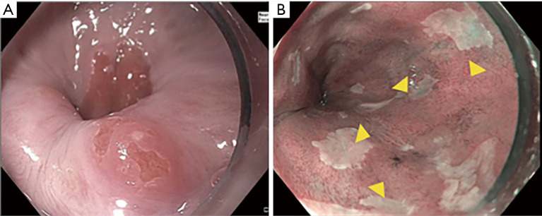
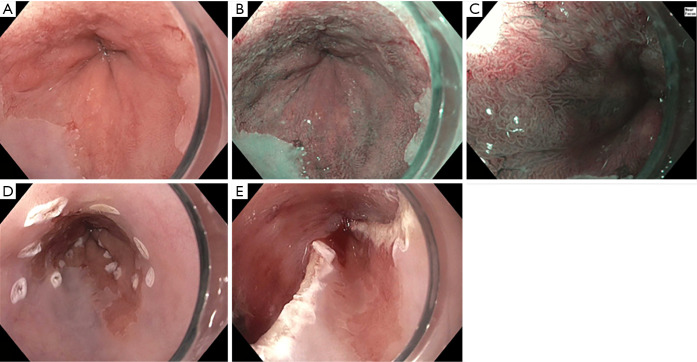
Esophageal adenocarcinoma (EAC) continues to be one of the fastest rising incident cancers in the Western population with the majority of patients presenting with late stage disease and associated with a dismal 5-year survival rate. Barrett's esophagus (BE) is the only identifiable precursor lesion to EAC. Strategies to screen for and survey BE are critical to detect earlier cancers and reduce morbidity and mortality related to EAC. A high-quality endoscopic examination with careful inspection of the Barrett's segment and adherence to the Seattle protocol for tissue sampling are critical. Advanced imaging modalities offer the potential to improve dysplasia detection, predict histopathology in real time and guide endoscopic eradication therapy (EET). Several technologies have been studied and although most are not yet recommended for routine clinical practice, high definition white light endoscopy (HD-WLE) as well as chromoendoscopy (including virtual chromoendoscopy) improved dysplasia detection in numerous studies supporting their use. Future studies should evaluate the role of artificial intelligence in optimizing detection of dysplasia in BE patients.
Keywords: Barrett’s esophagus (BE); Esophageal adenocarcinoma (EAC); advanced imaging; chromoendoscopy.
2021 Translational Gastroenterology and Hepatology. All rights reserved.
Conflict of interest statement
Conflicts of Interest: Both authors have completed the ICMJE uniform disclosure form (available at http://dx.doi.org/10.21037/tgh.2020.02.10). The series “Advanced Endoscopic Imaging of the GI Tract” was commissioned by the editorial office without any funding or sponsorship. JM Kolb received funding from the National Institutes of Health (NIH) T32-DK007038. S Wani is supported in part by the Department of Medicine Outstanding Early Scholars Program, and is a consultant for Medtronic, Boston Scientific, and Interspace.
Figures
References
-
- National Cancer Institute. SEER Cancer Statistics Review (CSR) 1975-2015. Available online: https://seer.cancer.gov/csr/1975_2015/
Publication types
Grants and funding
LinkOut - more resources
Full Text Sources
Other Literature Sources