Review
. 2021 Mar;383(3):931-947.
doi: 10.1007/s00441-020-03314-6. Epub 2021 Jan 6.

Structural aspects of the aging invertebrate brain

Affiliations
Review

Structural aspects of the aging invertebrate brain

Sandra C Koch et al. Cell Tissue Res. 2021 Mar.

Abstract

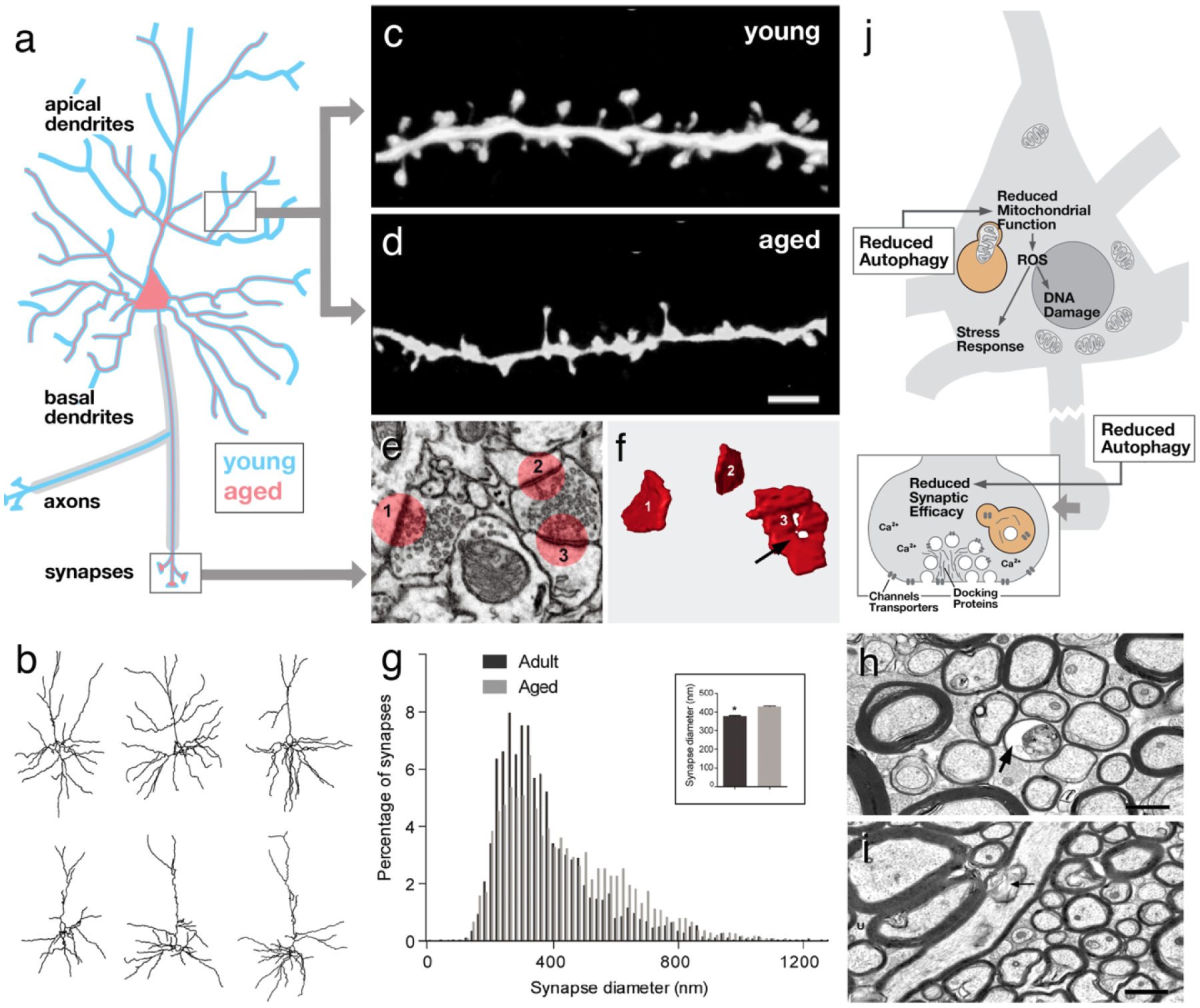
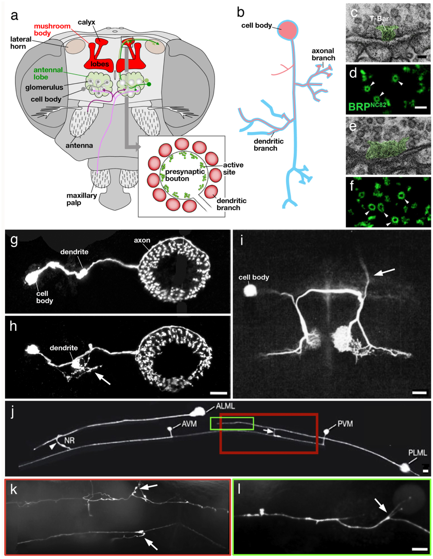
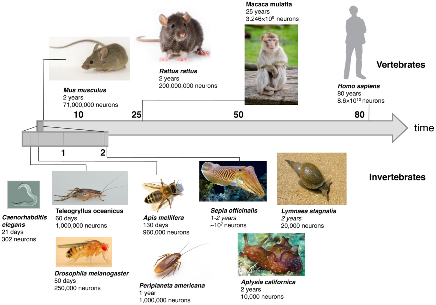
Aging is characterized by a decline in neuronal function in all animal species investigated so far. Functional changes are accompanied by and may be in part caused by, structurally visible degenerative changes in neurons. In the mammalian brain, normal aging shows abnormalities in dendrites and axons, as well as ultrastructural changes in synapses, rather than global neuron loss. The analysis of the structural features of aging neurons, as well as their causal link to molecular mechanisms on the one hand, and the functional decline on the other hand is crucial in order to understand the aging process in the brain. Invertebrate model organisms like Drosophila and C. elegans offer the opportunity to apply a forward genetic approach to the analysis of aging. In the present review, we aim to summarize findings concerning abnormalities in morphology and ultrastructure in invertebrate brains during normal aging and compare them to what is known for the mammalian brain. It becomes clear that despite of their considerably shorter life span, invertebrates display several age-related changes very similar to the mammalian condition, including the retraction of dendritic and axonal branches at specific locations, changes in synaptic density and increased accumulation of presynaptic protein complexes. We anticipate that continued research efforts in invertebrate systems will significantly contribute to reveal (and possibly manipulate) the molecular/cellular pathways leading to neuronal aging in the mammalian brain.

Keywords: Axons; Dendrites; Invertebrates; Neurons; Normal aging; Synapses.


Conflict of interest statement

Conflict of interest On behalf of all authors, the corresponding author states that there is no conflict of interest.

Figures

Fig. 1
Fig. 1
Structural aspects of aging in vertebrate neurons. a Schematic representation of cortical pyramidal neuron. Aged neuron (red outline), in a region-specific and cell type-specific manner, exhibits a reduction in terminal dendrites, often restricted to specific (e.g., apical) parts of their arbor. Loss of myelinated axons has also been recorded. b Three representative superficial pyramidal cells of mouse prefrontal cortex at the age of 8 months (top) and 28 months (bottom; from Grill and Riddle 2002, with permission). c, d At the synaptic level, postsynaptic dendritic spines frequently show a reduction in size and/or density with age (from Dickstein et al. 2013, with permission). e, f, g Whereas spines maybe decreased in size, individual presynaptic sites (i.e., active zones) can be increased (g) and show ultrastructural abnormalities, such as an enhanced fraction of ring-shaped zones (arrow in F; from Calì et al. 2018, with permission). h, i Myelinated axons of the cingulate bundle in 24-year-old macaque (h) are reduced in number and density compared to 9-year-old animal (i; from Bowley et al. 2010, with permission). Some aged fibers show ballooning of myelin sheath (arrow). j Schematic representation of neuron (inset: magnified view of synapse) summarizing cellular/molecular motifs associated with aging. Scale bars: 2 microns (c, d); 2 microns (h, i)
Fig. 2
Fig. 2
Structural aspects of aging in invertebrate neurons. a Olfactory pathway in Drosophila. Olfactory receptor neurons (magenta) project to the antennal lobe (green); from here olfactory projection neurons (green) relay the signal to the calyx of the mushroom body (red). Bulbous presynaptic boutons of projection neurons (see inset) contact dendritic branches of intrinsic mushroom body neurons (Kenyon cells; after Perisse et al. 2013, with permission). b Schematic representation of insect neuron, showing cell body, dendritic branches and axonal branches. Aged neuron (red outline) frequently lack dendritic and/or axonal branches; another characteristic is the occurrence of new branches at ectopic positions. c-f Ultrastructure of mushroom body calycal synapse in young adult fly (c, d) and aged fly (e, f). Note widening of T-bar (green shading), an ultrastructurally distinct feature of the active zone, in aged synapse (e). (d, f) show increased diameter of active zone, labeled by an antibody against the presynaptic protein BRP, in aged fly (f) compared to young fly (d; from Gupta et al. 2016, with permission). g, h GFP labeled ring neurons of the Drosophila ellipsoid body in young fly (g) and aged fly (h). Note ectopic fine branches sprouting from short bulbous dendrite (arrow). i Fluorescently labeled acoustic interneuron in cricket. Arrow points at ascending branch that is found in majority of young animals but absent from most aged animals (from Atkins and Pollack 1986; with permission). j-l Fluorescently labeled ALM and PLM touch receptor neurons in young C. elegans worm (j) and aged animal (k, l). Boxed regions in (j) corresponds to parts of fibers shown, for an aged animal, in (k) and (l). Note ectopic branches (arrows) occurring in aged animal (From Toth et al. 2012, with permission). Scale bars: 50 nm (c, e); 500 nm (d, f); 10 microns (g, h, j, k, l); 100 microns (i)
Fig. 3
Fig. 3
Overview of life span and neuron numbers of reviewed vertebrates and invertebrates. Vertebrates: mouse (Mus musculus), rat (Rattus rattus), monkey (Macaca mulatta) and human (Homo sapiens). Invertebrates: worm (Caenorhabditis elegans), cricket (Teleogryllus oceanicus), fruit fly (Drosophila melanogaster), honeybee (Apis mellifera), cockroach (Periplaneta Americana), cuttlefish (Sepia officinalis), sea hare (Aplysia caliornica) and pond snail (Lymnea stagnalis)

Similar articles

Cited by

References

    1. Akhmedov K, Rizzo V, Kadakkuzha BM, et al. (2013) Decreased Response to Acetylcholine during Aging of Aplysia Neuron R15. PLoS One 8:e84793. - PMC - PubMed
    1. Alexander AG, Marfil V, Li C (2014) Use of C. elegans as a model to study Alzheimer’s disease and other neurodegenerative diseases. Front Genet 5:1–21 - PMC - PubMed
    1. Alvarez J, Alvarez-Illera P, García-Casas P, et al. (2020) The Role of Ca2+ Signaling in Aging and Neurodegeneration: Insights from Caenorhabditis elegans Models. Cells 9:204 - PMC - PubMed
    1. Amdam GV. (2011) Social context, stress, and plasticity of aging. Aging Cell 10:18–27 - PubMed
    1. Atkins G, Pollack GS (1986) Age-dependent occurrence of an ascending axon on the omega neuron of the cricket, Teleogryllus oceanicus. J Comp Neurol 243:527–534 - PubMed